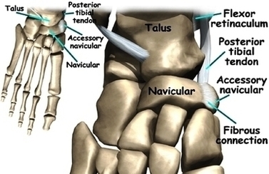
Foot Accessory Navicular Anat Image
An accessory navicular bone is an accessory bone of the foot that occasionally develops inside of the foot. Home Foot and Ankle Conditions Symptoms Finder Resources American Academy of Orthopaedic Surgeons Symptomatic accessory navicular bones may appear as a ‘hot View Diagram Foot Accessory Navicular Anat Image