Author Archives: anatomy
The Gender Of Child Determined At Weeks Of Pregnancy Image

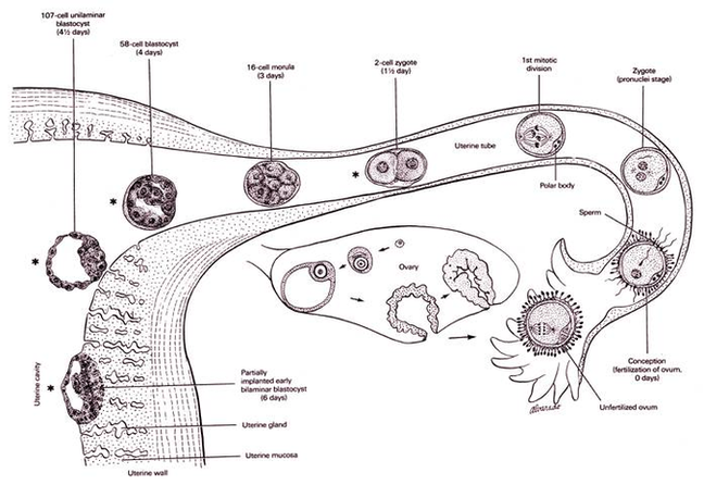
How a Baby’s XX Gender or XY Gender is Determined Females have an XX pair of sex chromosomes, and males, an XY pair. A baby’s gender is determined by the sperm cell that fertilizes a woman’s egg. Sperm carries one View Diagram The Gender Of Child Determined At Weeks Of Pregnancy Image
Diagram Crown Before After Image

Before: the patient had four existing crowns on the front teeth. She complained that the crowns were too white and also that her gums were swollen. The main reason for this, is that the crowns did not fit her teeth View Diagram Diagram Crown Before After Image
Http://www.dreamstime.com/ image61290
Stock photos are made by people just like you. Talented photographers and graphic artists upload their images to Dreamstime every day where those in need of great stock images can find them, purchase them, and use them. Our customers find View Diagram Http://www.dreamstime.com/ image61290
Diagram Wrist Anatomy Arteries Image
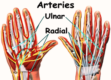
Wrist. The wrist connects the hand to the forearm. It consists of the distal ends of the radius and ulna bones, eight carpal bones, and the proximal ends of five metacarpal bones. This arrangement of bones allows for a wide View Diagram Diagram Wrist Anatomy Arteries Image
Blood Pressure Chart Example Image
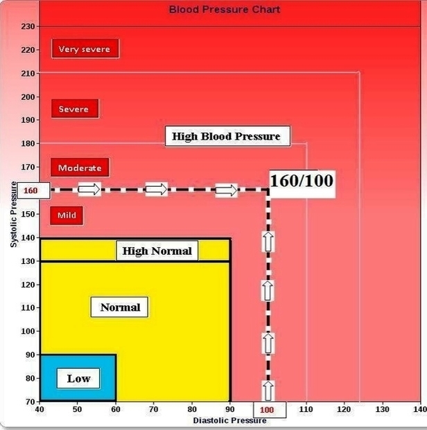
Log blood pressure readings and heart rate with this printable blood pressure chart template. This accessible blood pressure tracker template will generate a chart giving a visual representation of the data so you can analyze the readings over time. Blood View Diagram Blood Pressure Chart Example Image
Diabetes Diet Plan Image

The American Diabetes Association offers a simple method of meal planning. In essence, it focuses on eating more vegetables. Follow these steps when preparing your plate: Fill half of your plate with nonstarchy vegetables, such as spinach, carrots and tomatoes. View Diagram Diabetes Diet Plan Image
Cub Cells Lesson Figure Image
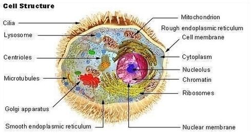
In the second lesson, students learn about the basics of cellular respiration. They also learn about the application of cellular respiration to engineering and bioremediation. The third lesson continues students’ education on cells in the human body and how (and View Diagram Cub Cells Lesson Figure Image
Synovial Joint Diagram Image
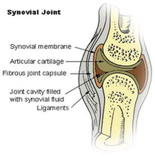
The structure of a synovial joint is demonstrated by a diagram in which the articulating bones are surrounded by the articular capsule, which comprises an exterior fibrous capsule and an interior synovial membrane. Start studying label the synovial joint. Learn View Diagram Synovial Joint Diagram Image
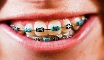
Dental Braces Image
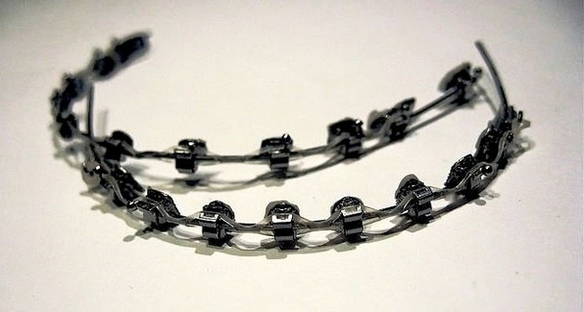
A macro shot of dental braces, (orthodontic braces) are a device used in orthodontics to align teeth and their position with Closing of gap with dental braces. Braces are orthodontic devices that straighten crooked and misaligned teeth. When people think View Diagram Dental Braces Image
Diagram Gasser Fig La Image
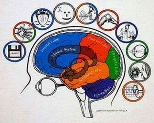
Brain Anatomy Shirt Large Image
Close Up Kid With Braces Image
Dental Implants Mesa Image
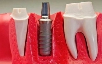
2,547 dental implant stock photos and images available, or search for dentures or dental implant surgery to find more great stock photos and pictures. Although the whole unit is usually referred to as a “dental implant,” the implant itself is View Diagram Dental Implants Mesa Image
Diabetes Diet Chart1 Image

How to optimize diabetic with a diet chart? Getting optimal results from a series of diabetic diet processes is the dream of everyone who does it. So an effort to use charts will be done. The way to get the View Diagram Diabetes Diet Chart1 Image
Diagram Anatomy Of Ear Image

12,612 human ear anatomy stock photos, vectors, and illustrations are available royalty-free. Picture of the Ear. The spiral-shaped cochlea is part of the inner ear; it transforms sound into nerve impulses that travel to the brain. The fluid-filled semicircular canals View Diagram Diagram Anatomy Of Ear Image
Diagram Foot Anatomy Bones Image
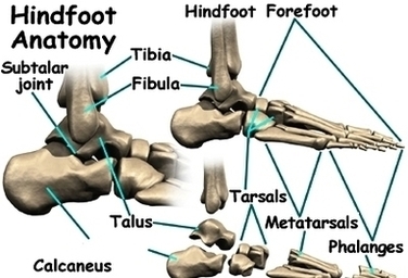
When one looks at the anatomy of the foot, they would realize that the foot has a complex mechanical and structural architecture. The ankle joint is the shock absorber of the foot. Apart from 28 bones, 33 joints, muscles, ligaments, View Diagram Diagram Foot Anatomy Bones Image
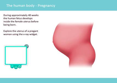
Diagram Getty Rm Of Week Fetus Image
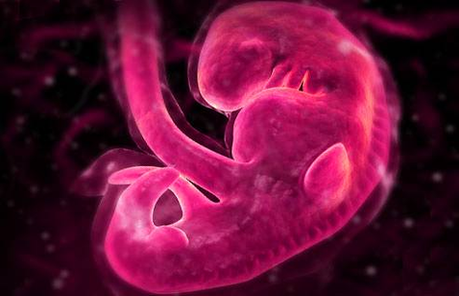
Diagram Of Anterior Leg Muscles Anterior View Madell Online Image
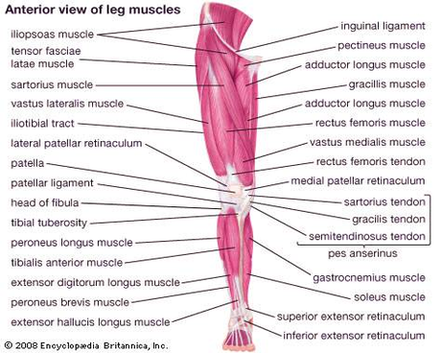
The anterior muscles of the leg. When studying the muscles of the leg, we can examine them by four primary groupings, which are the anterior, fibular/lateral, superficial posterior and deep posterior compartments. These are defined by intermuscular septa and surrounded View Diagram Diagram Of Anterior Leg Muscles Anterior View Madell Online Image
Diagram Of Baae Ac Beb Image