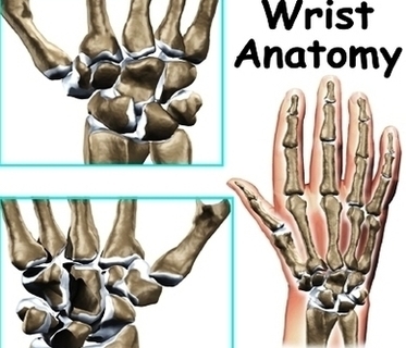
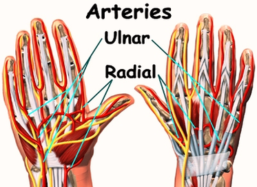
Diagram Wrist Anatomy Arteries Image
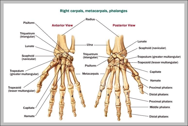
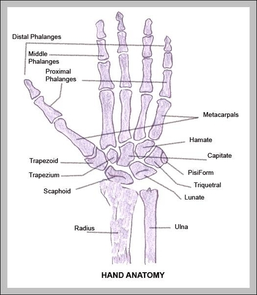
Wrist. The wrist connects the hand to the forearm. It consists of the distal ends of the radius and ulna bones, eight carpal bones, and the proximal ends of five metacarpal bones. This arrangement of bones allows for a wide View Diagram Diagram Wrist Anatomy Arteries Image