Foot Diagram With Labels Image

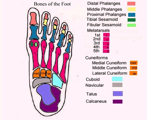
Foot Diagram: Labeled Anatomy. The foot diagram has a complex structure made up of bones, ligaments, muscles, and tendons. Understanding the structure of the foot is best done by looking at a foot diagram where the anatomy has been labeled. If you would like to learn all the parts of the foot structure, you have come to the right place.

2,592 foot diagram stock photos, vectors, and illustrations are available royalty-free. See foot diagram stock video clips

In the diagram above, you can see all of the bones of the foot clearly labeled. By now you should be familiar enough with the names, shapes and locations of these bones that you can label them based on the diagram image alone. Are you up to the challenge? Below you will find an unlabelled diagram PDF of the foot bones, ready for you to fill out.

Foot Diagram With Labels Image Diagram - Chart - diagrams and charts with labels. This diagram depicts Foot Diagram With Labels Image

Foot Diagram With Labels Image

Screening With Colonos Sm Image

As such, “screening” describes a colonoscopy that is routinely performed on an asymptomatic person for the purpose of testing for the presence of colorectal cancer or colorectal polyps. Whether a polyp or cancer is ultimately found does not change the screening intent of that procedure.

E/M Service Prior to a Screening Colonoscopy Typically, procedure codes with 0, 10 or 90-day global periods include pre-work, intraoperative work, and post-operative work in the Relative Value Units (RVUs) assigned. As a result, CMS’ policy does not allow for payment of an Evaluation and Management (E/M) service prior to a screening colonoscopy.

Colonoscopy is one of the most sensitive tests currently available for colon cancer screening. The doctor can view your entire colon and rectum. Abnormal tissue, such as polyps, and tissue samples (biopsies) can be removed through the scope during the exam.

Screening With Colonos Sm Image Diagram - Chart - diagrams and charts with labels. This diagram depicts Screening With Colonos Sm Image

Screening With Colonos Sm Image

Px Tubal Pregnancy With Embryo Image

Tubal pregnancy: A pregnancy that is not in the usual place within the uterus but is located in the Fallopian tube. Tubal pregnancies are due to the inability of the fertilized egg to make its way through the Fallopian tube into the uterus.

Tubal pregnancies are most often diagnosed in the sixth through the ninth weeks of pregnancy. 5 Your healthcare provider will be able to diagnose a tubal pregnancy after a medical exam and other testing, which may include: 6 Pelvic examination to test for pain, tenderness, or a mass in the abdomen

Pregnancy tests taken at the beginning of a tubal pregnancy will usually show a positive result, even though the pregnancy won’t be able to grow into a healthy baby. Some people may experience no symptoms at first, but as the fertilized egg continues to grow in the fallopian tube, there can be some serious and more noticeable signs, including: 2

Px Tubal Pregnancy With Embryo Image Diagram - Chart - diagrams and charts with labels. This diagram depicts Px Tubal Pregnancy With Embryo Image

Px Tubal Pregnancy With Embryo Image

Anatomy Of The Chicken With Text Image

The leg of the chicken is similar to the human anatomy except that the hip bone is fused with the backbone. This provides a strong and rigid union in conjunction with powerful muscles. We have covered much of the hens’ reproductive system before, but we are going to do a brief review here to refresh memories. Why do birds lay eggs?

The basic external parts of a chicken include the comb, beak, wattles, ears, earlobes, eyes, eye rings, wings, tail, thighs, hocks, shanks, spurs, claws and toes. As Figures 1 and 2 show, both male and female chickens have these basic parts. The differences between males and females include the size of the comb and wattles, …

The chicken has flexible bones within its backbone and neck. There are a total of 39 bones within a chicken’s spine. This helps protect the skull and gives a chicken the ability to turn their heads 180 degrees. The largest bone within the bird’s body is the sternum. This bone covers half of a bird’s body.

Anatomy Of The Chicken With Text Image Diagram - Chart - diagrams and charts with labels. This diagram depicts Anatomy Of The Chicken With Text Image

Anatomy Of The Chicken With Text Image