Anatomy Leg By Bk Image

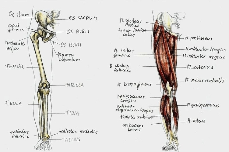
4,236 human leg anatomy stock photos and images available, or start a new search to explore more stock photos and images.

When your non-health science friends talk about the ‘leg’, they mean the entire lower extremity. However, in the world of anatomy, the ‘leg’ strictly means the portion between the knee and the ankle joints. It is composed of several structures: In this page, we will take a look at all of the above as well as the anatomy of the knee.

The lower leg contains two major long bones, the tibia and the fibula, which are both very strong skeletal structures. The tibia (also called the shinbone) is located near the midline of the leg and is the thicker and stronger of the two bones.

Anatomy Leg By Bk Image Diagram - Chart - diagrams and charts with labels. This diagram depicts Anatomy Leg By Bk Image

Anatomy Leg By Bk Image

Anatomy Leg By Bk1 Image

72,510 leg anatomy stock photos, vectors, and illustrations are available royalty-free.

When your non-health science friends talk about the ‘leg’, they mean the entire lower extremity. However, in the world of anatomy, the ‘leg’ strictly means the portion between the knee and the ankle joints. It is composed of several structures: In this page, we will take a look at all of the above as well as the anatomy of the knee.

The leg is divided into three major anatomical compartments, each one containing a specific group of muscles: 1 anterior group 2 lateral group 3 posterior group

Anatomy Leg By Bk1 Image Diagram - Chart - diagrams and charts with labels. This diagram depicts Anatomy Leg By Bk1 Image

Anatomy Leg By Bk1 Image

Diagram Of Anterior Leg Muscles Anterior View Madell Online Image

The anterior muscles of the leg. When studying the muscles of the leg, we can examine them by four primary groupings, which are the anterior, fibular/lateral, superficial posterior and deep posterior compartments. These are defined by intermuscular septa and surrounded by the deep fascia of the leg.

Tibialis anterior muscle This muscle is the most anterior and medial of all four anterior leg muscles. It originates from the proximal portion of the leg, precisely, from the lateral tibial condyle and proximal half of the tibial shaft, in addition to the adjacent portion of the interosseous membrane.

Do you visualise all of the muscles from the thigh down to the ankle? In fact, the muscles of the leg refer to the muscles found in the region between the knee and foot . In this article, we’re going to be teaching you about every last one of them, including their individual names, origins, insertions, innervations and functions.

Diagram Of Anterior Leg Muscles Anterior View Madell Online Image Diagram - Chart - diagrams and charts with labels. This diagram depicts Diagram Of Anterior Leg Muscles Anterior View Madell Online Image

Diagram Of Anterior Leg Muscles Anterior View Madell Online Image

Leg Workout Routine Leg Anatomy Image

How to Design a Leg Workout Using the 15 Best Exercises. 1 1. Back squat. Target your posterior chain — or the back of your body, including the glutes and hamstrings — with a back squat. 2 2. Front squat. 3 3. Romanian deadlift. 4 4. Good mornings. 5 5. Walking lunges. More items

As a beginner looking to increase general fitness — and following the theme of keeping things simple — choose 3–5 exercises per leg workout. Then complete 3 sets of 8–12 reps of each exercise, ensuring that you’re working your muscles to fatigue but not failure (1). Why no more than 5 exercises?

When it comes to designing an effective leg workout, simpler is better. The basic lower body movements — squats, hip hinges (deadlifts), and lunges — should comprise the majority of your programming. These movements inherently focus on the major muscle groups of the legs: the glutes, quads, hamstrings, and calves.

Leg Workout Routine Leg Anatomy Image Diagram - Chart - diagrams and charts with labels. This diagram depicts Leg Workout Routine Leg Anatomy Image

Leg Workout Routine Leg Anatomy Image

Anatomy Of The Lower Leg Achilles Tendon Image

The Achilles tendon is also located in the lower leg. The lower leg is made up of two very strong, long bones: the tibia and the fibula. The tibia, also known as the shin bone, is the stronger and larger of the two. It is located toward the middle of the lower leg. The fibula, or calf bone, is smaller and is located on the outside of the lower leg.

The Achilles tendon attaches the muscles of the calves to the bones of the ankle and foot. The ankle is a joint that connects the lower leg to the foot.

Pain in the back of the heel, worse with shoes on, is the common symptom. Physical examination: To look for Achilles tendon problems, an examiner checks for pain, swelling, warmth, thickening, or discoloration around the heel and leg. A knot on the back of the leg may be present with Achilles tendon rupture.

Anatomy Of The Lower Leg Achilles Tendon Image Diagram - Chart - diagrams and charts with labels. This diagram depicts Anatomy Of The Lower Leg Achilles Tendon Image

Anatomy Of The Lower Leg Achilles Tendon Image

Power Leg Muscle Group Image

Leg muscles are bundles of fibrous tissue that contract and relax to exert forces on bones and move the legs The main muscle groups in the legs are: quadriceps, hamstrings, adductors in the upper leg or thigh, and the calves in the lower legs.

The legs are the lower extremities that allow for standing, walking, running, and more. Their movement is produced by the contraction and relaxation of leg muscles and their connection to the skeleton through tendons. The movement of the legs produced by these muscles happens around the leg joints, particularly the hips, knees and ankles.

171,684 leg muscle stock photos, vectors, and illustrations are available royalty-free.

Power Leg Muscle Group Image Diagram - Chart - diagrams and charts with labels. This diagram depicts Power Leg Muscle Group Image

Power Leg Muscle Group Image

Anatomy Of The Leg Muscles Image

72,510 leg anatomy stock photos, vectors, and illustrations are available royalty-free.

Leg muscles are bundles of fibrous tissue that contract and relax to exert forces on bones and move the legs The main muscle groups in the legs are: quadriceps, hamstrings, adductors in the upper leg or thigh, and the calves in the lower legs.

The legs are the lower extremities that allow for standing, walking, running, and more. Their movement is produced by the contraction and relaxation of leg muscles and their connection to the skeleton through tendons. The movement of the legs produced by these muscles happens around the leg joints, particularly the hips, knees and ankles.

Anatomy Of The Leg Muscles Image Diagram - Chart - diagrams and charts with labels. This diagram depicts Anatomy Of The Leg Muscles Image

Anatomy Of The Leg Muscles Image

Anatomy Of The Posterior Upper Leg Hamstrings Image

The hamstring muscles, or simply the hamstrings, are a group of three long muscles located in the posterior compartment of the thigh, shaping up the surface anatomy of this region. These muscles are the biceps femoris, semimembranosus and semitendinosus muscles.

Posterior thigh muscles (Hamstring muscles) The hamstring muscles, or simply the hamstrings, are a group of three long muscles located in the posterior compartment of the thigh, shaping up the surface anatomy of this region. These muscles are the biceps femoris, semimembranosus and semitendinosus muscles.

398 hamstring anatomy stock photos and images available, or start a new search to explore more stock photos and images. Posterior View Of The Human Right Leg, Showing The Muscles Of The Hip, Thigh, And Lower Leg, As Well As The Tibial Nerve.

Anatomy Of The Posterior Upper Leg Hamstrings Image Diagram - Chart - diagrams and charts with labels. This diagram depicts Anatomy Of The Posterior Upper Leg Hamstrings Image

Anatomy Of The Posterior Upper Leg Hamstrings Image

Diagram Leg Bones Anatomy Image

72,510 leg anatomy stock photos, vectors, and illustrations are available royalty-free.

The legs include the upper leg, knee, lower leg, ankle, and foot. (The hip is part of the trunk of the body as opposed to the leg so that joint and its muscles will not be covered in this lesson)

The bones of the lower leg are the tibia and fibula. The tibia bears most of the weight, and the fibula serves as attachment points for the lower leg muscles. The two tibialis and three peroneus muscles are also found in the lower leg

Diagram Leg Bones Anatomy Image Diagram - Chart - diagrams and charts with labels. This diagram depicts Diagram Leg Bones Anatomy Image

Diagram Leg Bones Anatomy Image

Leg Xray Image

1,659 leg xray stock photos and images available, or search for broken leg xray or dog broken leg xray to find more great stock photos and pictures.

A leg X-ray can detect broken bones, assist a physician in setting the broken bone, and can monitor the treatment process to determine whether the bone is properly aligned and the break is healing properly.

Medical doctor woman holds x-ray of leg beside patient. Two doctors discussing a patient X Ray on the screen showing a broken leg.They discuss images and notes on the mobile phone. Broken leg, X-ray. vet doctor watching x-ray image of dog which jumped from sofa and broke leg. Young doctor examining a leg X-ray and pointing out a part of it.

Leg Xray Image Diagram - Chart - diagrams and charts with labels. This diagram depicts Leg Xray Image

Leg Xray Image

Veins And Arteries Of The Leg Image

8,053 veins and arteries stock photos and images available, or search for blood vessel or human veins to find more great stock photos and pictures.

Important veins of the leg include the internal and external iliac veins, femoral vein, saphenous vein, popliteal vein, tibial vein, and the venous arch of the foot. Nerves in the leg send messages to the brain, including indications of heat, pain, and movement.

This propels blood up the leg until it can empty into large veins like the femoral vein and then travel back to the heart via the iliac veins and inferior vena cava. The superficial veins are also influenced by the musculevenous pump although not to the extent as the deep veins.

Veins And Arteries Of The Leg Image Diagram - Chart - diagrams and charts with labels. This diagram depicts Veins And Arteries Of The Leg Image

Veins And Arteries Of The Leg Image