The 3D images of the wrist joint and carpal bones are three-dimensional reconstructions obtained from a scanner. 340 anatomical structures of the wrist were labeled, accessible on “Anatomical parts”: General anatomy: the different regions of the wrist and hand (carpal, metacarpal, hypothenar and thenar eminence).
Anatomy, medical imaging and e-learning for healthcare professionals – IMAIOS Anatomy atlas e-Anatomy and medical imaging learning resources available on the web and mobile (Android and iOS) ×Your email address is not verified.
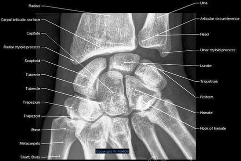
Plain frontal and side-view X-Rays of the wrist show the lower extremities of the radius and ulna, the radiocarpal joint, the carpal bones (scaphoid, capitate, trapezium, trapezoid, hamate, lunate, pisiform and triquetral) and the carpometacarpal joints. Anatomy atlas , Radiographs , Wrist : Anterior-posterior view
Wrist Radiography Ap View Imaios Anatomy En Medical Image

