

Tag Archives: illustration
Diagram Prenatal Exercise Illustration Image

Ear Anatomy Illustration Image
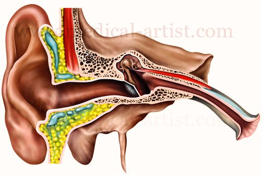
12,612 human ear anatomy stock photos, vectors, and illustrations are available royalty-free. In most mammals, the visible ear is a flap of tissue that is also called the pinna or the auricle. Vertebrates have a pair of ears, placed somewhat View Diagram Ear Anatomy Illustration Image
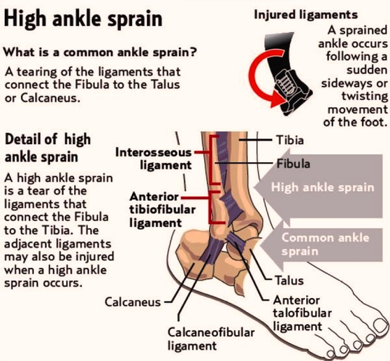
High Ankle Sprain Illustration Photos Image
Asthma Attack Illustration Image
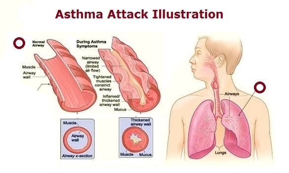
Diagram Of Human Anatomy Mediastinum Left View Illustration Large Image
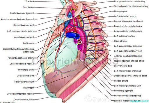
Nerves: details the nervous anatomy of the mediastinum including notably the route of the phrenic and vagus nerves as well as the anatomy of the sympathetic trunk at the thoracic level. Heart: the external morphology of the heart in the View Diagram Diagram Of Human Anatomy Mediastinum Left View Illustration Large Image
Muscles Arm Human Anatomy Illustration Diagram En Medical Image
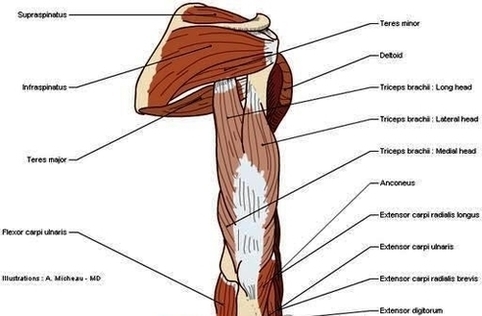
The human arm is divided into two main regions, the portion from the elbow to the wrist known as the forearm, and the segment from the shoulder to the elbow referred to as the arm. Arm muscle anatomy enables the View Diagram Muscles Arm Human Anatomy Illustration Diagram En Medical Image