Category Archives: Anatomy
Shoulder Anatomy Intro Image
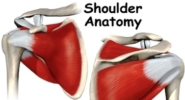
The Anatomy of the Shoulder. The acromioclavicular joint is where the acromion, part of the shoulder blade (scapula) and the collar bone (clavicle) meet. The glenohumeral joint is where the ball (humeral head) and the socket (the glenoid) meet. The View Diagram Shoulder Anatomy Intro Image
Muscles Stability Image
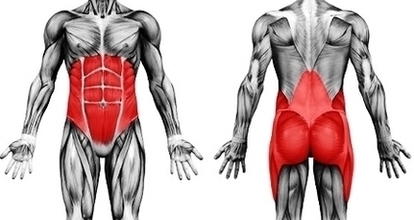
Following are the five major muscles / muscle groups of sacroiliac stabilization that should be assessed and likely worked with manual therapy when the client presents with a sacroiliac joint condition. Piriformis Gluteus Maximus (superior deep fibers) Coccygeus and Levator View Diagram Muscles Stability Image
Blood Flow Diagram Image
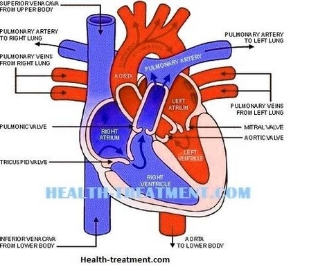
The blue arrows represent the flow of deoxygenated blood through the right side of the heart. The red arrows represent the flow of oxygenated blood through the left side of the heart. Diagram: Blue arrows demonstrate flow of deoxygenated blood View Diagram Blood Flow Diagram Image
Cavities Diagram Image
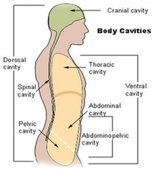
38 nasal cavity diagram stock photos and images available or start a new search to explore more stock photos and images. Many sinuses, which are small, hollow holes, connect to the nasal cavity. Oral cavity : also known as the View Diagram Cavities Diagram Image
Screening Type Fy Image

Elements Molecular Neurobiology Image
High Blood Pressure Hypertophy Heart Anatomy Model Image

There is still much uncertainty about the pathophysiology of hypertension. A small number of patients (between 2% and 5%) have an underlying renal or adrenal disease as the cause for their raised blood pressure. In the remainder, however, no clear View Diagram High Blood Pressure Hypertophy Heart Anatomy Model Image
Diagram Shoulder Blade Pain Image

If there is no evidence of an injury, it may be either referred pain or pain from a damaged nerve. The most common causes of chronic shoulder pain are strains and sprains from overuse or overexertion of a shoulder. Why View Diagram Diagram Shoulder Blade Pain Image
Diabetes Diet Images Image

Muscles In Back Diagram Image
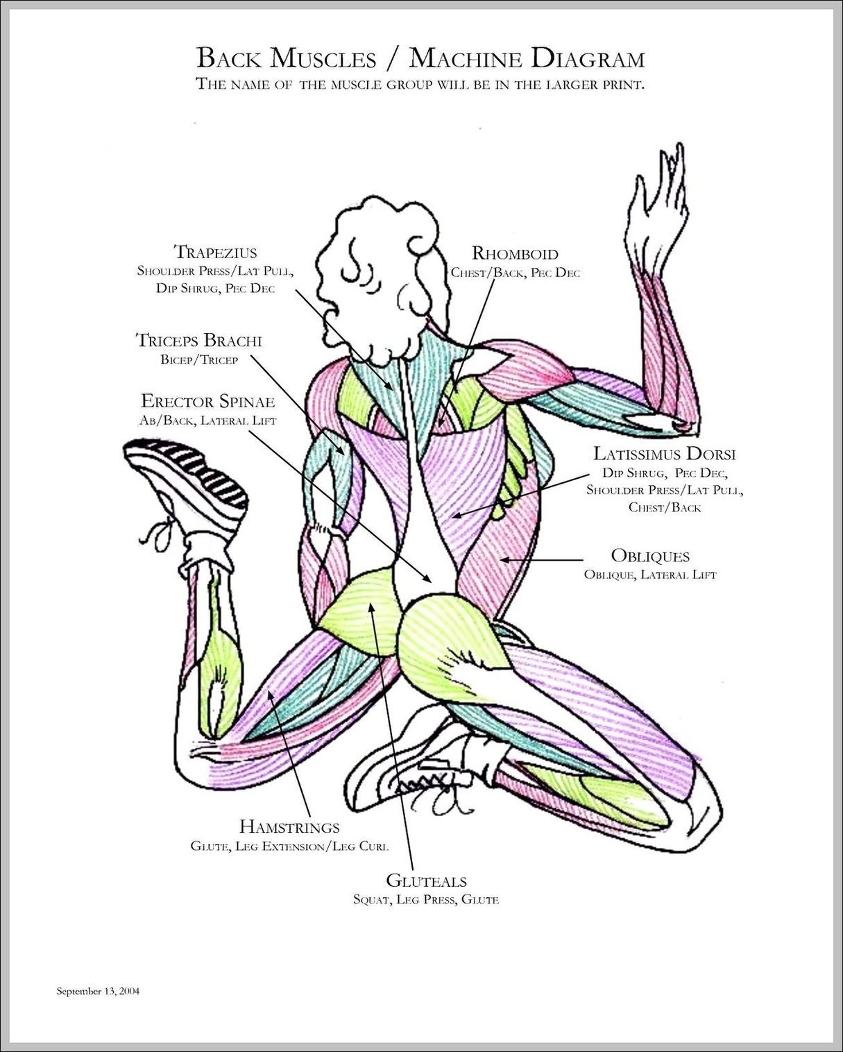
25,649 back muscle anatomy stock photos, vectors, and illustrations are available royalty-free. There are three different muscle groups found in the back: the superficial group, the deep group, and the intermediate group. Muscles found in the superficial group include rhomboid View Diagram Muscles In Back Diagram Image
Diabetic Food Pyramid Figure Image
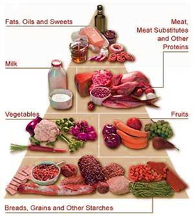
A food pyramid for diabetes could help you to understand and comprehend what this looks like in daily life. The glycemic indexrepresents the magnitude of the increase in blood glucose that occurs after ingestion of the food. This index measures View Diagram Diabetic Food Pyramid Figure Image
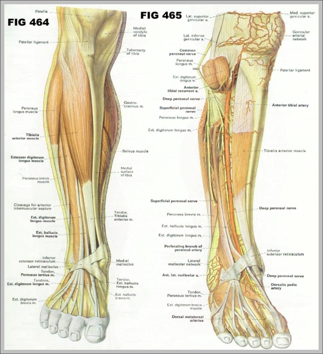
Anatomy Of The Leg Muscles Image
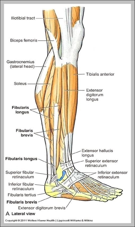
72,510 leg anatomy stock photos, vectors, and illustrations are available royalty-free. Leg muscles are bundles of fibrous tissue that contract and relax to exert forces on bones and move the legs The main muscle groups in the legs are: quadriceps, View Diagram Anatomy Of The Leg Muscles Image
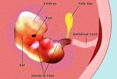
Pregnancy Weeks Pregnant Embryo Fetus Development Photos Image
Pregnancy Weeks Pregnant Fetus Development Diagram Image
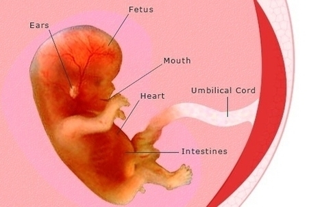
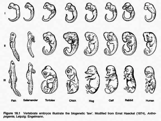
Your baby moves through different stages, starting as a blastocyst, then maturing into an embryo, and then a fetus. Around the five week mark, your baby’s heart will begin to beat, at 27 weeks they’ll have regular sleep and wake View Diagram Pregnancy Weeks Pregnant Fetus Development Diagram Image
Function Of The Fallopian Tubes Image
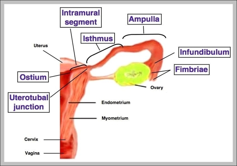
The fallopian tubes are also known as oviducts or uterine tubes. They are important parts of the female reproductive system. Fertilization normally happens in the fallopian tubes. Medically reviewed by Healthline’s Medical Network on March 25, 2015. The uterine tube View Diagram Function Of The Fallopian Tubes Image
Colorectal Cancer Lg1 Image
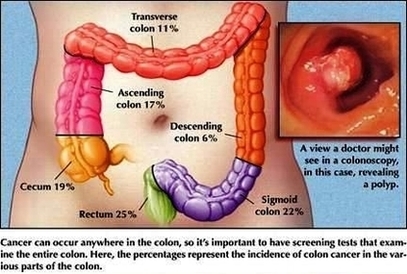
4,035 colorectal cancer stock photos and images available, or search for colon cancer screening or colonoscopy to find more great stock photos and pictures. Doctor goes over a patient”s x-ray, screening for colon cancer. There is no single cause of View Diagram Colorectal Cancer Lg1 Image
Calf Muscle Anatomy Image
Knee Anatomy Ligaments Image
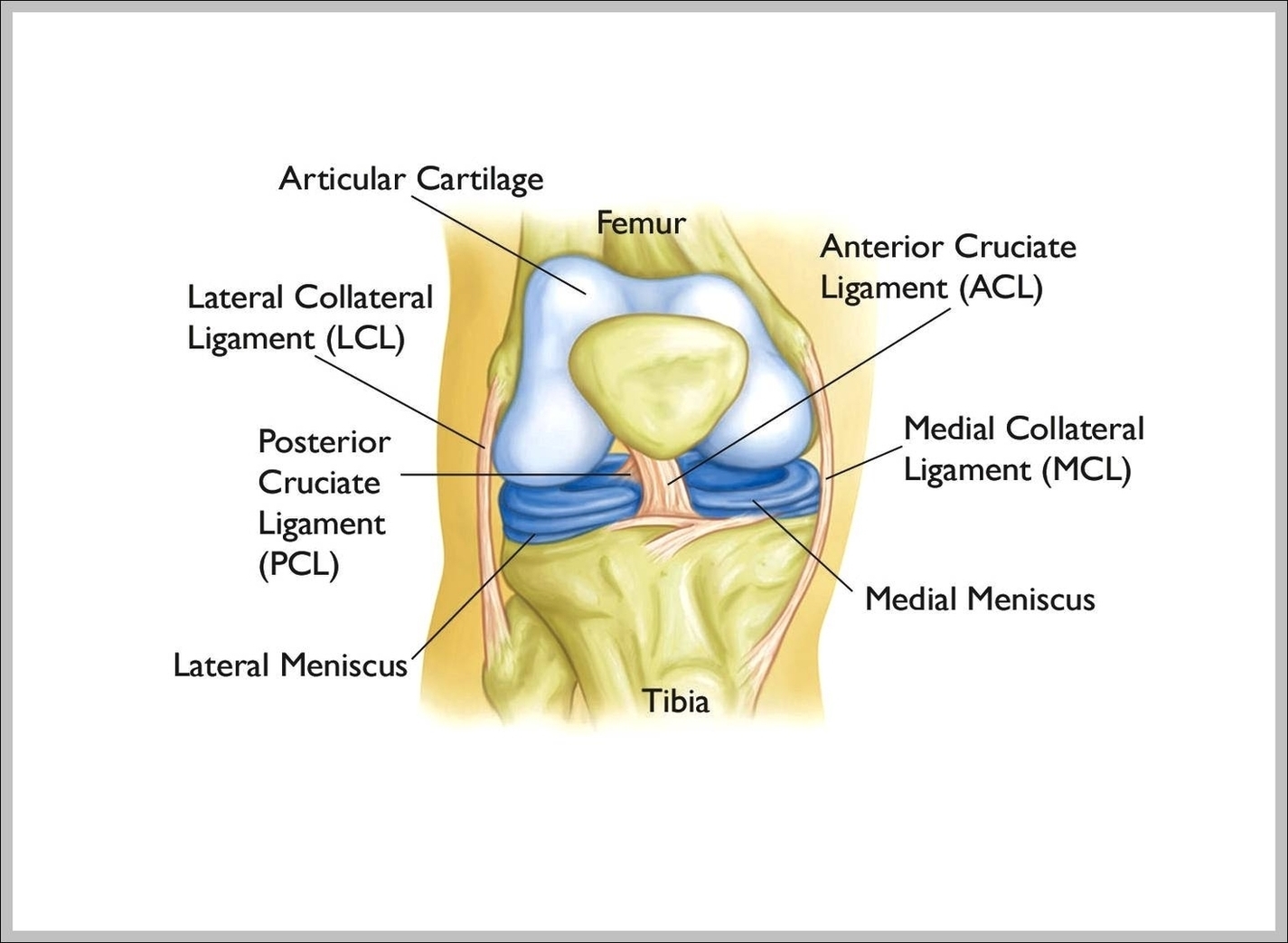
The patella (kneecap) sits over the front of the knee joint. Four major ligaments connect the bones and stabilize the knee joint . In the image above, the physician is pointing to the anterior cruciate ligament, or ACL, one of View Diagram Knee Anatomy Ligaments Image
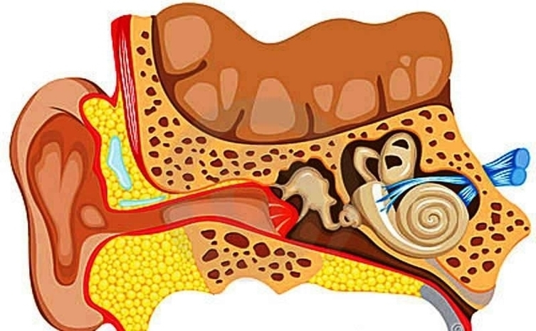
Human Ear Anatomy Image