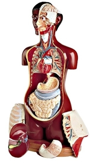
Human Muscle Model Image

105,188 human muscle anatomy stock photos, vectors, and illustrations are available royalty-free.

Forearms – Anatomy Muscles Rhomboid minor and rhomboid major, levator scapulae and latissimus dorsi muscles – didactic board of anatomy of human bony and muscular system, posterior view Abs – Anatomy Muscles Leg muscles of the man Human Body Organs (Lungs) 3D Triceps – Anatomy Muscles Hand muscle connection with brain

Last Updated: Jul 16, 2019 The muscular system is responsible for the movement of the human body. Attached to the bones of the skeletal system are about 700 named muscles that make up roughly half of a person’s body weight. Each of these muscles is a discrete organ constructed of skeletal muscle tissue, blood vessels, tendons, and nerves.

Human Muscle Model Image Diagram - Chart - diagrams and charts with labels. This diagram depicts Human Muscle Model Image

Human Muscle Model Image

Diagram Model Embryo To Cell Stage Icon Image

The image of the embryo or the egg in the mother’s womb has a reddish tint. human embryo development stock pictures, royalty-free photos & images The image of the embryo or the egg in the mother’s womb has a… Stages of embryo development Stages of embryo development. Vector flat infographic icons. human embryo development stock illustrations

Mosaic image composed of 9 sections. Also known as a blastocyst where the inner cell mass or embryoblast forms the embryo. The outer layer of cells are the trophoblast. human embryo development stock pictures, royalty-free photos & images Fertilized, high quality, human egg after five days just prior to transfer into the woman’s uterus.

1,727 human embryo development stock photos and images available, or search for ivf or ivf process to find more great stock photos and pictures. Vector human embryo development circle flat icon Vector human embryo development circle with female uterus icon.

Diagram Model Embryo To Cell Stage Icon Image Diagram - Chart - diagrams and charts with labels. This diagram depicts Diagram Model Embryo To Cell Stage Icon Image

Diagram Model Embryo To Cell Stage Icon Image

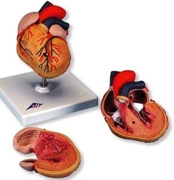
High Blood Pressure Hypertophy Heart Anatomy Model Image

There is still much uncertainty about the pathophysiology of hypertension. A small number of patients (between 2% and 5%) have an underlying renal or adrenal disease as the cause for their raised blood pressure. In the remainder, however, no clear single identifiable cause is found and their condition is labelled “essential hypertension”.

In response to this pressure overload inside the heart, heart muscle cells may respond by getting thicker along the inner walls of the heart. This thickened wall can cause the left chamber to weaken, stiffen and lose elasticity, which may prevent healthy blood flow.

Left ventricular hypertrophy is more common in people who have uncontrolled high blood pressure. But no matter what your blood pressure is, developing left ventricular hypertrophy puts you at higher risk of a heart attack and stroke. Treating high blood pressure can help ease your symptoms and may reverse left ventricular hypertrophy.

High Blood Pressure Hypertophy Heart Anatomy Model Image Diagram - Chart - diagrams and charts with labels. This diagram depicts High Blood Pressure Hypertophy Heart Anatomy Model Image

High Blood Pressure Hypertophy Heart Anatomy Model Image

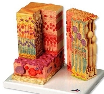
Microscohuman Eye Anatomy Model Retina Image

162 retina diagram stock photos and images available, or start a new search to explore more stock photos and images. Color diagram depicting retinal bands and retinal detachment following a puncture wound to the eye, from the volume “Atlas and epitome of… Diagram Of A Retinal Detachment.

A schematic section through the human eye with a schematic enlargement of the retina The retina is approximately 0.5 mm thick and lines the back of the eye.

Much more information concerning pathology of the whole eye and retina can be found in a website made by eye pathologist Dr. Nick Mamalis, Moran Eye Center. Fig. 22. A view of the fundus of the eye and of the retina in a patient who has age-related macular degeneration. Fig. 23.

Microscohuman Eye Anatomy Model Retina Image Diagram - Chart - diagrams and charts with labels. This diagram depicts Microscohuman Eye Anatomy Model Retina Image

Microscohuman Eye Anatomy Model Retina Image