Knee Anatomy Ligaments Image

This human anatomy diagram with labels depicts and explains the details and or parts of the Knee Anatomy Ligaments Image. Human anatomy diagrams and charts show internal organs, body systems, cells, conditions, sickness and symptoms information and/or tips to ensure one lives in good health.

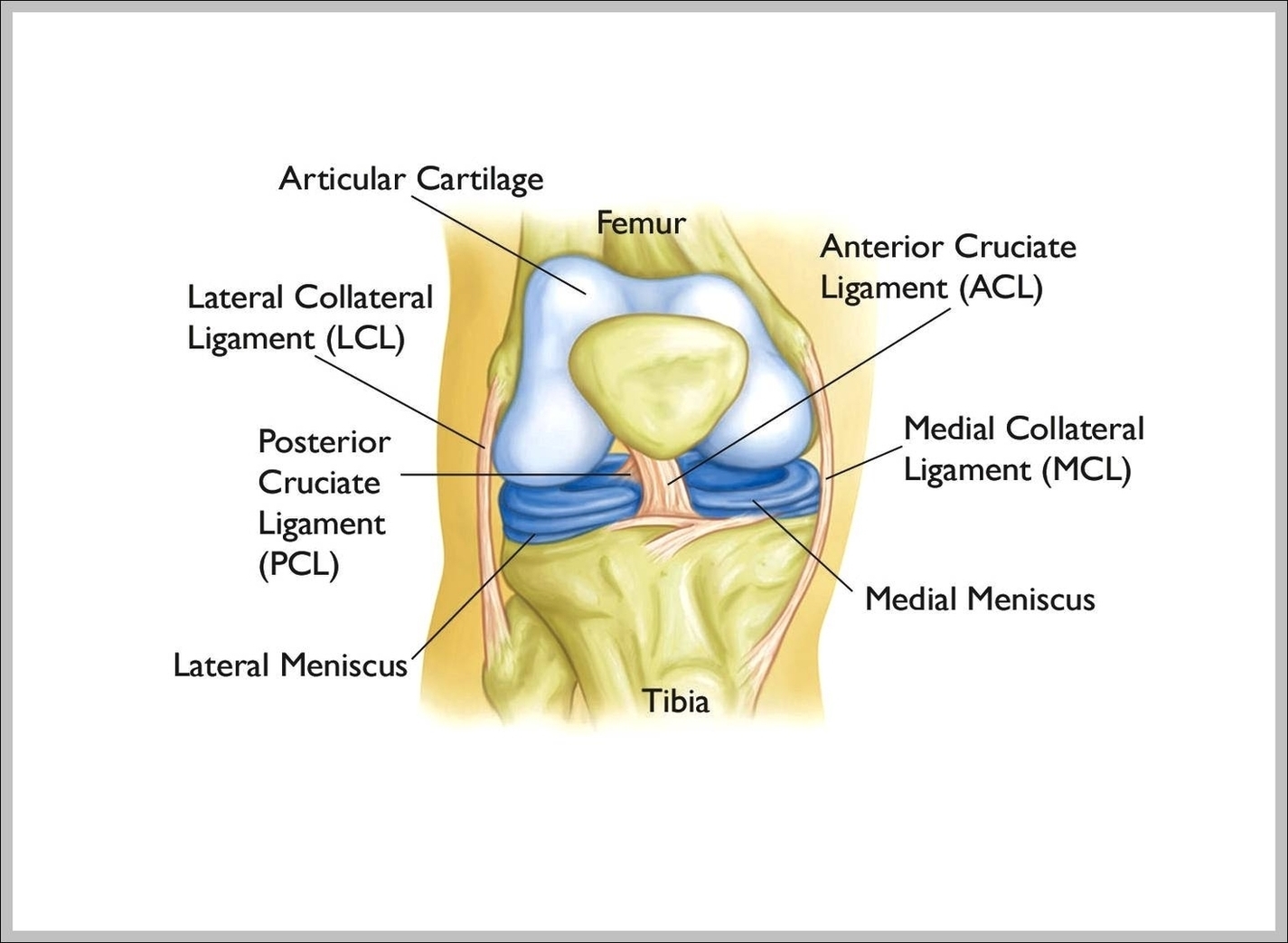
The patella (kneecap) sits over the front of the knee joint. Four major ligaments connect the bones and stabilize the knee joint . In the image above, the physician is pointing to the anterior cruciate ligament, or ACL, one of these important ligaments. Inside the knee joint is a smooth cover on the ends of the bone called articular cartilage.

1,274 knee ligament stock photos and images available, or search for acl or biomedical illustration to find more great stock photos and pictures. Orthopedic surgery, Saint George Clinic, Nice, France, Treating a torn cruciate ligament using the Sambba technique, carried out under arthroscopy,…

The normal human anatomy of a knee, anterolateral view. Shown are the patella, anterior cruciate ligament, patellar ligament, fibular collateral ligament, lateral meniscus, lateral femoral condyle, femur, tibia, and fibula. Anterior cruciate ligament “A doctor, pointing at anterior cruciate ligament of a right knee.

Knee Anatomy Ligaments Image

Knee Anatomy Ligaments Image


Tags: ,