Picture Of Vertebrae 2 Image

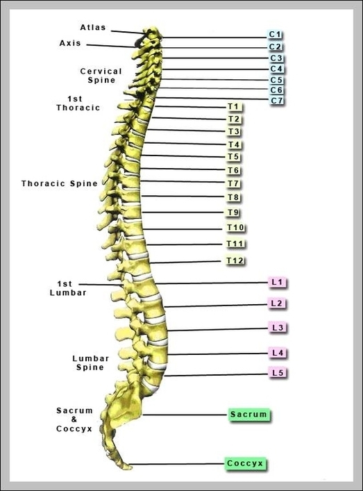
11,423 spine vertebrae stock photos and images available or start a new search to explore more stock photos and images.

The spine is a darker white than the background, and shows all of the intricate details of the various parts from the top to the bottom. The spine is curved and shows spaces in between the vertebrae. human vertebra stock pictures, royalty-free photos & images The human spine on a white background.

Vector diagram for medical, educational and science use human vertebra stock illustrations Spinal cord. sections of vertebral column: cervical, thoracic,… Spinal cord. sections of vertebral column: cervical, thoracic, and lumber spine, sacrum and coccyx. Human silhouette with backbone, intervertebral discs, hip bones and joints.

Picture Of Vertebrae 2 Image Diagram - Chart - diagrams and charts with labels. This diagram depicts Picture Of Vertebrae 2 Image

Picture Of Vertebrae 2 Image

Testis 2 Diagram Image

What are testes? The testes — also called testicles — are two oval-shaped organs in the male reproductive system. They’re contained in a sac of skin called the scrotum. The scrotum hangs outside the body in the front of the pelvic region near the upper thighs.

The testes (testicles) are male reproductive glands found in a saccular extension of the anterior abdominal wall called the scrotum. They are in ovoid shape, sized four to six centimeters in length. Testes develop retroperitoneally on the posterior abdominal wall and descend to scrotum before birth.

Each testis is divided by invaginations of the tunica albuginea that divide it into several hundred small segments called lobules. Each lobule contains several tightly coiled tubes called seminiferous tubules. The walls of the seminiferous tubules contain the germ cells, Sertoli cells, and Leydig cells that give the testes their function.

Testis 2 Diagram Image Diagram - Chart - diagrams and charts with labels. This diagram depicts Testis 2 Diagram Image

Testis 2 Diagram Image

Ovary 2 Diagram Image

114 ovary diagram stock photos and images available, or start a new search to explore more stock photos and images. Generalized Flower With Parts ; Diagram Showing Arrangement Of Floral Parts In Cross Section At The Flower’S Base .

Structure of the Ovary. The ovaries are almond-shaped structures located on either side of the uterus, and closely related to several anatomical structures in the pelvic region. Each ovary has average dimensions of 3 cm (length), 2 cm (breadth), and 1 cm (thickness); hence being smaller than its male analogue – the testis.

The ovary is a ductless reproductive gland in which the female reproductive cells are produced. Females have a pair of ovaries, held by a membrane beside the uterus on each side of the lower abdomen.

Ovary 2 Diagram Image Diagram - Chart - diagrams and charts with labels. This diagram depicts Ovary 2 Diagram Image

Ovary 2 Diagram Image

Terminal Ileum Anatomy 2 Image

Terminal Ileum. The terminal ileum is the distal end of the small intestine that intersects with the large intestine.

Ileum (“twisted intestine”) is the final and the longest portion of the small intestine, the ileum measures about 3 m (9.8 ft ) and contributes to almost 60% of the length of the small intestine. The ileum joins the large intestine at a smooth muscle sphincter called the ileocecal sphincter (valve).

Characteristics & Features. The transition from the jejunum to the ileum is not sharply marked, while at the distal end, the ileum opens into the large intestine. At the junction between the ileum and the caecum lies the ileocecal valve (ileal ostium), a functional sphincter formed by the circular muscle layers of both the ileum and cecum.

Terminal Ileum Anatomy 2 Image Diagram - Chart - diagrams and charts with labels. This diagram depicts Terminal Ileum Anatomy 2 Image

Terminal Ileum Anatomy 2 Image

Picture Of Body Organs Location 2 Image

12,251 human body organs anatomy stock photos and images available, or start a new search to explore more stock photos and images.

Human body internal organs and parts info poster vector. Heart and brain, liver and kidneys. Thymus gland and reproductive system of male and female

Woman anatomy cardiovascular system with skeleton and internal rear and front views. An illustration depicting the major organs in the human body, as well as a ghosted view of the bones of the skeleton. Full length view of male human body with organs.

Picture Of Body Organs Location 2 Image Diagram - Chart - diagrams and charts with labels. This diagram depicts Picture Of Body Organs Location 2 Image

Picture Of Body Organs Location 2 Image