Cavities Diagram Image

This human anatomy diagram with labels depicts and explains the details and or parts of the Cavities Diagram Image. Human anatomy diagrams and charts show internal organs, body systems, cells, conditions, sickness and symptoms information and/or tips to ensure one lives in good health.

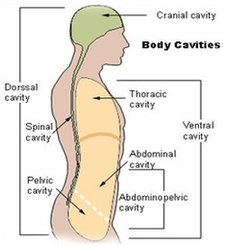
38 nasal cavity diagram stock photos and images available or start a new search to explore more stock photos and images.

Many sinuses, which are small, hollow holes, connect to the nasal cavity. Oral cavity : also known as the mouth. Holds the teeth, the gums, the tongue, the roof of the mouth, and floor of the mouth. Orbital Cavities : there are two orbital cavities, each containing the eyeball and nerves, blood vessels, and muscles related to the eye.

Ventral Cavity The ventral cavity is located at the front of the trunk and contains the thoracic cavity and the abdominopelvic cavity. The thoracic cavity and abdominopelvic cavity are separated by the diaphragm, which is a thin skeletal muscle. Thoracic cavity : located in the chest (upper part of the trunk) and contains the heart and the lungs.

Cavities Diagram Image

Cavities Diagram Image


Tags: