Blood Flow Diagram Image

This human anatomy diagram with labels depicts and explains the details and or parts of the Blood Flow Diagram Image. Human anatomy diagrams and charts show internal organs, body systems, cells, conditions, sickness and symptoms information and/or tips to ensure one lives in good health.

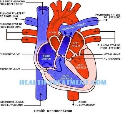
The blue arrows represent the flow of deoxygenated blood through the right side of the heart. The red arrows represent the flow of oxygenated blood through the left side of the heart. Diagram: Blue arrows demonstrate flow of deoxygenated blood through the right side of the heart.

Blood flows through the heart in the following order: 1) body –> 2) inferior/superior vena cava –> 3) right atrium –> 4) tricuspid valve –> 5) right ventricle –> 6) pulmonary arteries –> 7) lungs –> 8) pulmonary veins –> 9) left atrium –> 10) mitral or bicuspid valve –> 11) left ventricle –> 12) aortic valve –> 13) aorta –> 14) body.

The blood is pumped into the blood vessels through the heart. This pumping of blood goes through a complex process, where in the deoxygenated blood or impure blood is cleansed and oxygenated blood or pure blood is sent throughout the body.

Blood Flow Diagram Image

Blood Flow Diagram Image


Tags: ,