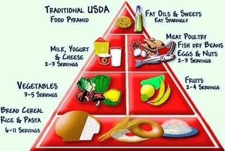
Category Archives: Anatomy
Diagram Diabetes Type Image
It is characterized by disturbances in carbohydrate, protein, or fat metabolism. It is classified as Type 1 (Insulin dependent or juvenile- onset diabetes) and Type 2 (Non- insulin dependent or also called as insulin- resistant disease). Insulin, a hormone released View Diagram Diagram Diabetes Type Image
Diagram Blood Pressure Chart Image
Blood pressure chart: What your reading means. By Mayo Clinic Staff. This blood pressure chart can help you figure out if your blood pressure is at a healthy level or if you’ll need to take some steps to improve your View Diagram Diagram Blood Pressure Chart Image
Dental Crowns Lake County Il1 Image
Dental Bridges Costa Rica Image
Hip Joint Muscles Image
hip joint anatomy images 19,596 hip joint anatomy stock photos, vectors, and illustrations are available royalty-free. See hip joint anatomy stock video clips Muscles of the Hip. The hip joint is one of the most flexible joints in the entire View Diagram Hip Joint Muscles Image
Back Shoulder Muscles Image
124,600 shoulder muscles stock photos, vectors, and illustrations are available royalty-free. See shoulder muscles stock video clips Other muscles that aid in shoulder movement include: 1 Infraspinatus: This rotator cuff muscle helps with the raising and lowering of the upper View Diagram Back Shoulder Muscles Image
Female Anatomy Diagram For Kids Image
465 female anatomy diagram stock photos and images available, or start a new search to explore more stock photos and images. Figures illustrating the difference between a natural waist and a constricted waist deformed through the use of corsets, from View Diagram Female Anatomy Diagram For Kids Image
Anatomy And Physiology Pictures Image
87,967 professional anatomy stock photos available royalty-free. Old vintage anatomy charts of the human body. Showing the skeletal system and various muscles, four figures in a row in different orientations SPINE Pain – Male Hurt Backbone isolated on white – View Diagram Anatomy And Physiology Pictures Image
Shoulder Muscles Anatomy Diagram Image
Based on their location, the shoulder muscles are grouped into: This page will introduce you to the anatomy and function of the shoulder muscles. Muscles of the shoulder are a group of muscles surrounding the shoulder joint, which move and View Diagram Shoulder Muscles Anatomy Diagram Image
Food Pyramid Fruits Image
Studio photo taken with DSRL Canon EOS 5D Mk II food pyramid stock pictures, royalty-free photos & images A large group of food. The assortment includes fruits, vegetables, meat, fish and dairy products. The food is mostly sorted by type. View Diagram Food Pyramid Fruits Image
Cervical Discectomy Rationale Image
Cervical discectomy is surgery to remove one or more discs from the neck. The disc is the pad that separates the neck vertebrae; ectomy means to take out. Usually a discectomy is combined with a fusion of the two vertebrae View Diagram Cervical Discectomy Rationale Image
Ankle Sprains Diagram Image
Bruising and swelling are common signs of a sprained ankle. If there is severe tearing of the ligaments, you might also hear or feel a “pop” when the sprain occurs. Most sprained ankles occur in the lateral ligaments on the View Diagram Ankle Sprains Diagram Image
Diagram Vintage Human Anatomy Muscles Print Rfecfcceedf Vmd Byvr Image
Spine anatomy poster shows 3 views of the spine, cervical, thoracic and luimbar vertebrae with cross-section of vertebral unit. Muscles and articulations of the human body. 15 labeled illustrations with cross sections of the shoulder, elbow and knee. The poster View Diagram Diagram Vintage Human Anatomy Muscles Print Rfecfcceedf Vmd Byvr Image
Rinc Of Fetus At Weeks Image Image
Diagram Of Tooth Implant Procedure Image
To understand how a dental implant procedure is performed, you should know the parts of the dental implant. A conventional dental implant consists of 3 parts: the implant body, abutment, and dental prosthesis. The implant body: it is similar in View Diagram Diagram Of Tooth Implant Procedure Image
Anatomy Of The Human Eye Cross Section View Image
Eye – Cross Section. A human eyeball cross section showing the following structures: iris, anterior limiting membrane, posterior limiting membrane, tendon of Eye anatomy, Human eye cross section physiology, Model of cornea and lens for ophthalmologist. Eye anatomy, Human eye View Diagram Anatomy Of The Human Eye Cross Section View Image
How To Become A Nurse Image
Stages Of Periodontitis Image
Periodontal disease is broken up into four separate stages: gingivitis, slight periodontal disease, moderate periodontal disease, and advanced periodontal disease. Gingivitis. Gingivitis is the only stage of periodontal disease that is reversible as it has not yet had time to View Diagram Stages Of Periodontitis Image
Human Body Muscle Diagram Image
43,556 human body diagram stock photos, vectors, and illustrations are available royalty-free. 128,412 muscle anatomy stock photos, vectors, and illustrations are available royalty-free. Last Updated: Jul 16, 2019 The muscular system is responsible for the movement of the human body. View Diagram Human Body Muscle Diagram Image