Cervical Discectomy Rationale Image

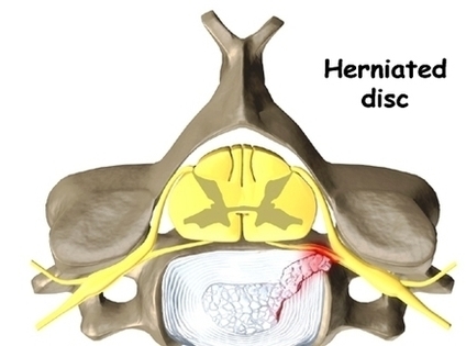
Cervical discectomy is surgery to remove one or more discs from the neck. The disc is the pad that separates the neck vertebrae; ectomy means to take out. Usually a discectomy is combined with a fusion of the two vertebrae that are separated by the disc. In some cases, this procedure is done without a fusion.

Peer Reviewed Anterior cervical discectomy and fusion (ACDF) is a type of neck surgery that involves removing a damaged disc to relieve spinal cord or nerve root pressure and alleviate corresponding pain, weakness, numbness, and tingling.

The surgery is approached through the anterior, or front, of the cervical spine (neck). The disc is then removed from between two vertebral bones. Fusion. A fusion surgery is done at the same time as the discectomy operation in order to stabilize the cervical segment.

Cervical Discectomy Rationale Image Diagram - Chart - diagrams and charts with labels. This diagram depicts Cervical Discectomy Rationale Image

Cervical Discectomy Rationale Image

Cervical Disk Mri Image

Magnetic resonance imaging (MRI) is a safe, painless test that uses radio waves and energy from strong magnets to create detailed images of your body. A cervical MRI scans the soft tissues of your neck and cervical spine. The cervical spine is the portion of your spine that runs through your neck.

Cervical MRI Scan 1 Magnetic resonance imaging (MRI) is a safe,… 2 An MRI scan uses a powerful magnetic field and radio waves to take detailed,… 3 A cervical spine MRI is usually used to diagnose the cause of neck pain. 4 MRI scans are very safe.

An MRI of the cervical spine does not need special preparations(11). However, individuals taking the MRI exam should remove metal objects, like eyeglasses and jewelry, as they can produce a bright or blank spot on the diagnostic film. Braces and dental fillings do not interfere with the procedure.

Cervical Disk Mri Image Diagram - Chart - diagrams and charts with labels. This diagram depicts Cervical Disk Mri Image

Cervical Disk Mri Image

Referral Multifidi Mid Cervical Image

Cervical multifidus muscles arise from the superior articular processes of C4 – C7. They extend superomedially to insert on the lateral aspect and the tips of the spinous processes of C2 – C5 vertebrae. Thoracic multifidus originate from the transverse processes of the thoracic vertebrae.

One study conducted by Bogduk (1996) identified the pain referral patterns of cervical facet joints. This information was derived from 61 volunteers who underwent a zygopophyseal joint injection and facet joint denervation in a pain clinic in Japan. They developed this referral chart for clinical use…… (Bogduk, 1996, p. 81).

As shown in the diagrams below, the multifidi trigger points lie along each side of the spine and sacrum, within the paravertebral canals. The diagrams above also show the referred pain associated with the multifidi trigger points.

Referral Multifidi Mid Cervical Image Diagram - Chart - diagrams and charts with labels. This diagram depicts Referral Multifidi Mid Cervical Image

Referral Multifidi Mid Cervical Image

Cervical Cancer Screening Bchjzgid Image

Widely used guidelines on screening women for cervical cancer have several important changes, including a recommendation to start screening at a slightly older age and use of an HPV test as the primary screening test. Credit: iStock. Routine cervical cancer screening is very effective for preventing cervical cancer and deaths from the disease.

Screening tests help find cervical cancer before any symptoms develop. When cervical cancer is found and treated early, the chances of successful treatment are better. A procedure in which cells are scraped from the cervix (the lower, narrow part of the uterus, or womb) and examined under a microscope.

A Pap test is a simple screening test that can detect cell changes in the cervix that may lead to cancer before women feel any symptoms. When to Get Screened. The Ontario Cervical Screening Program recommends that women who are or have been sexually active have a Pap test every 3 years starting at age 21.

Cervical Cancer Screening Bchjzgid Image Diagram - Chart - diagrams and charts with labels. This diagram depicts Cervical Cancer Screening Bchjzgid Image

Cervical Cancer Screening Bchjzgid Image