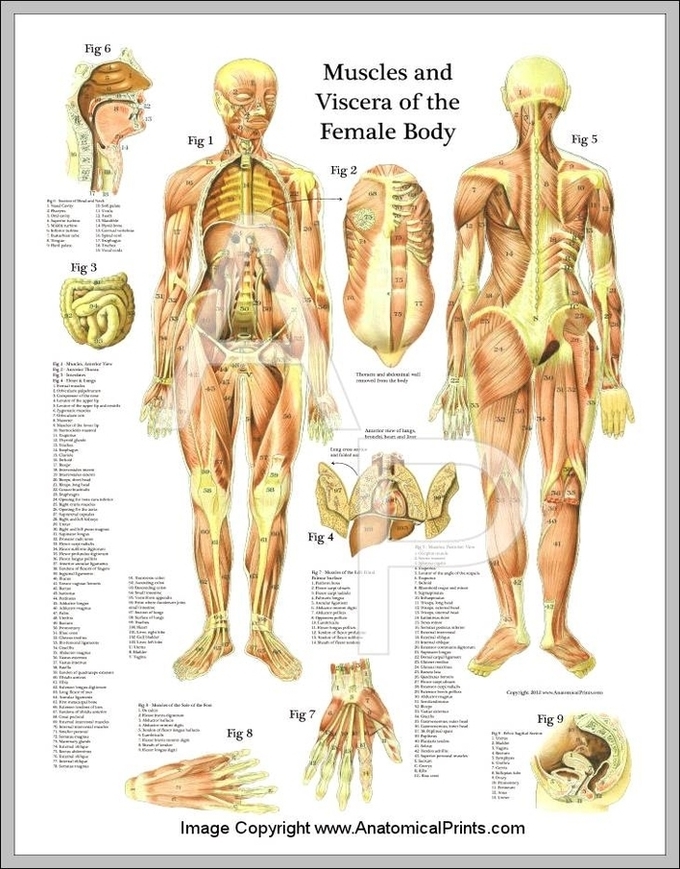
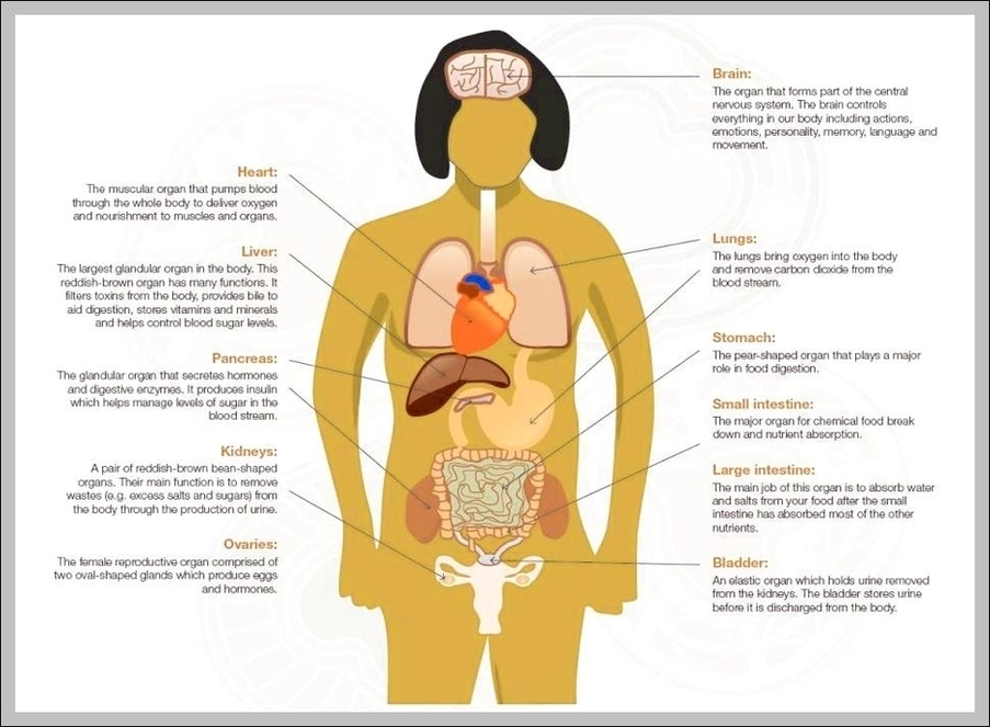
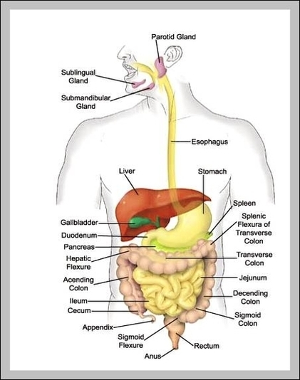
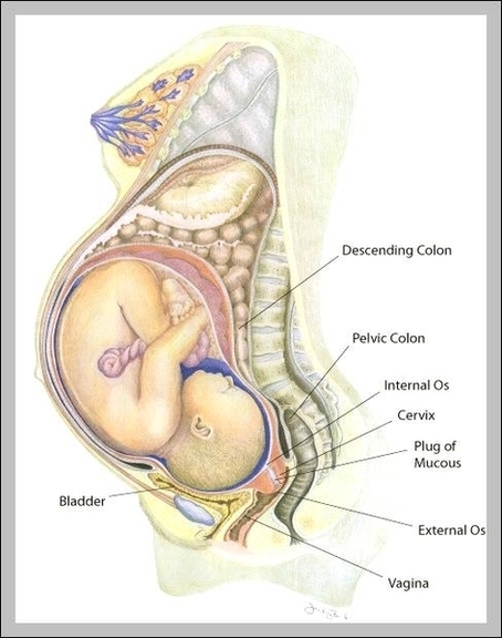
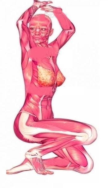
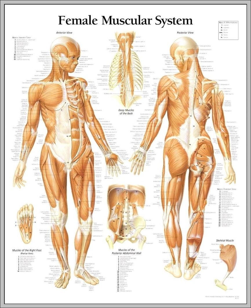
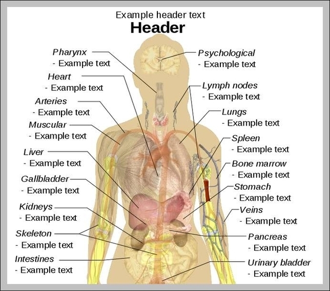
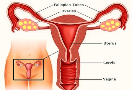
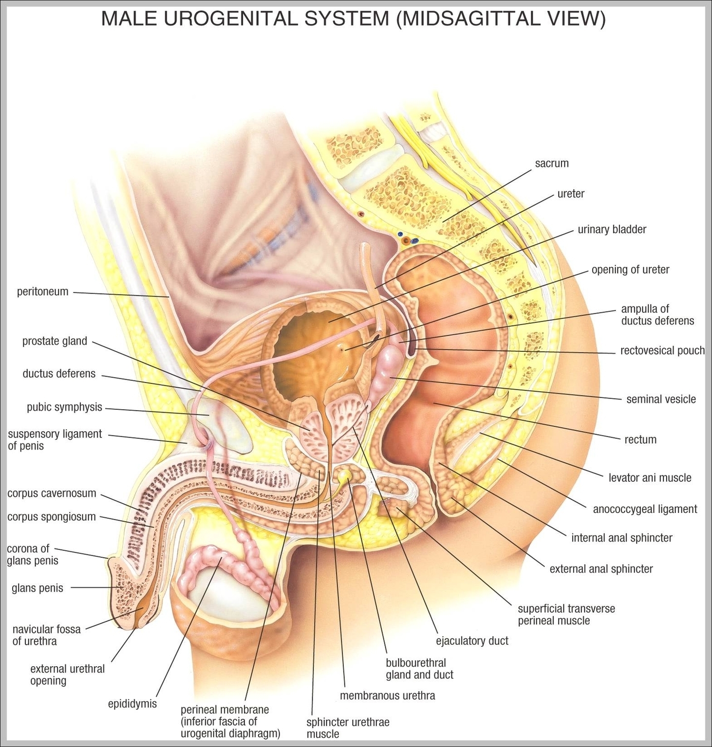
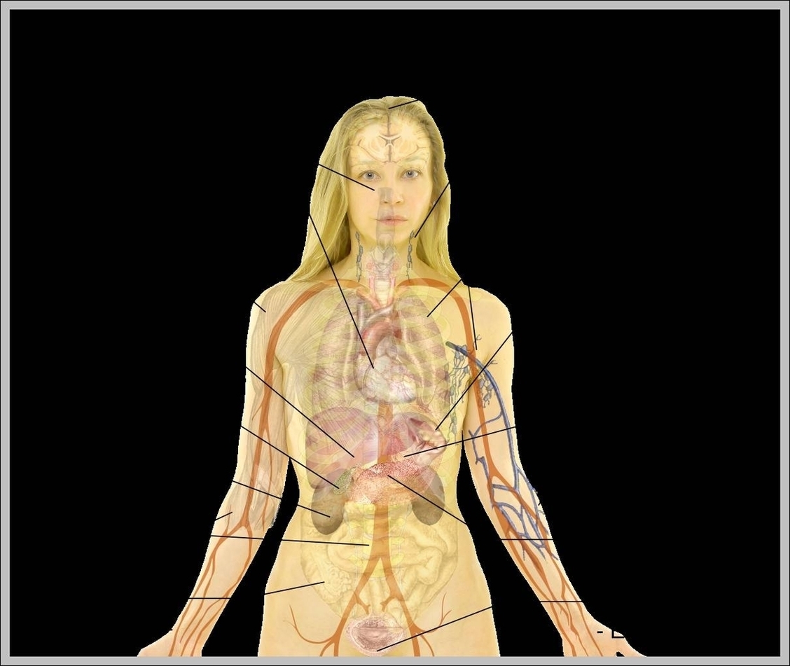
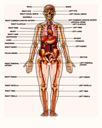
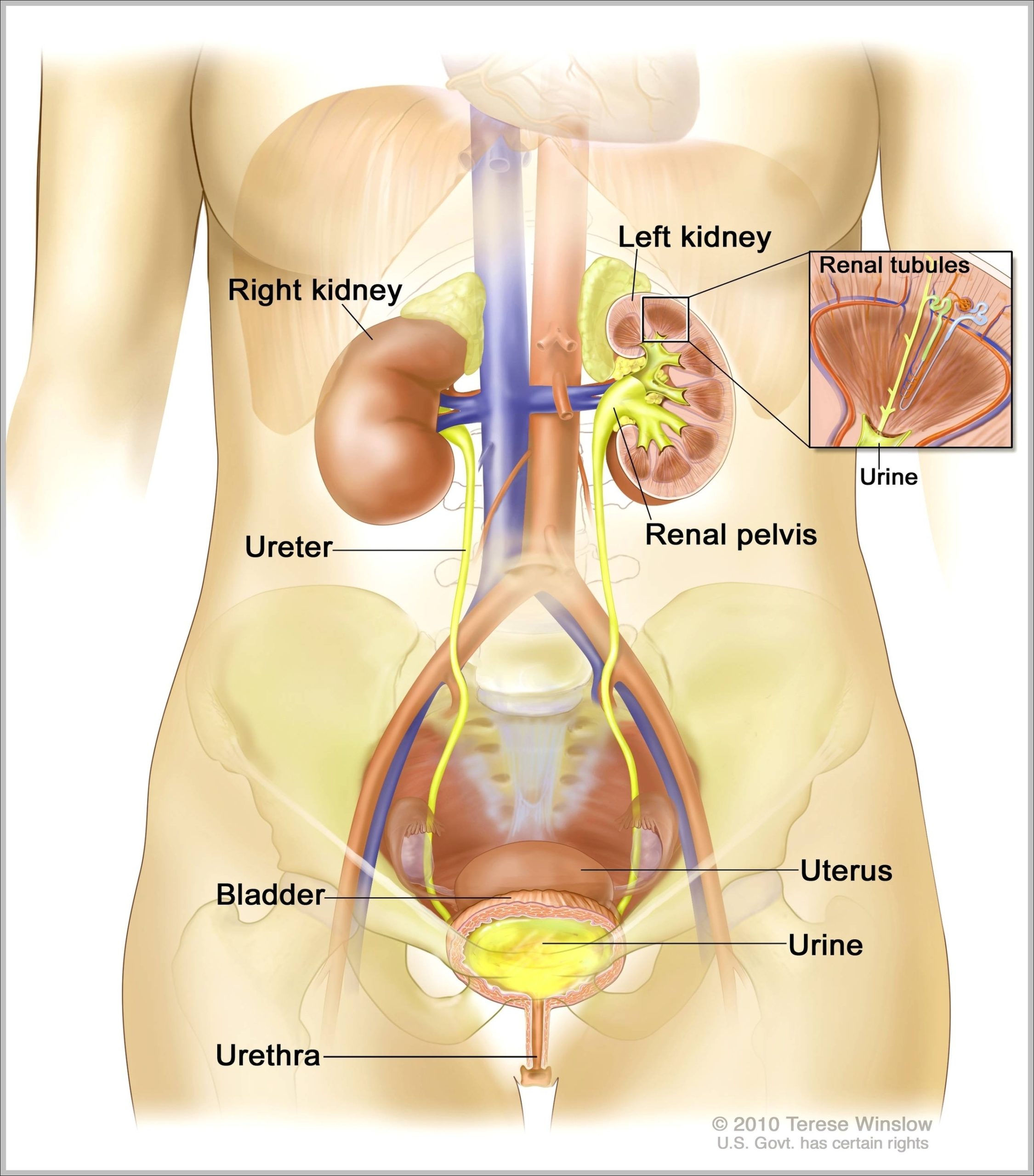
Female Muscle Anatomy Chart Image Diagram - Chart - diagrams and charts with labels. This diagram depicts Female Muscle Anatomy Chart Image
Tag Archives: female
Female Anatomy Diagram For Kids Image
465 female anatomy diagram stock photos and images available, or start a new search to explore more stock photos and images. Figures illustrating the difference between a natural waist and a constricted waist deformed through the use of corsets, from a published essay by…
Sexual anatomy that’s typically called female includes the vulva and internal reproductive organs like the uterus and ovaries. The vulva is the part of your genitals on the outside of your body — your labia, clitoris, vaginal opening, and the opening to the urethra (the hole you pee out of).
In This Section. Sexual anatomy that’s typically called female includes the vulva and internal reproductive organs like the uterus and ovaries. The vulva is the part of your genitals on the outside of your body — your labia, clitoris, vaginal opening, and the opening to the urethra (the hole you pee out of).
Female Anatomy Diagram For Kids Image Diagram - Chart - diagrams and charts with labels. This diagram depicts Female Anatomy Diagram For Kids Image
Internal Female Anatomy Pictures Image
Anatomy Of Female Image
24,838 female anatomy stock photos and images available or search for female anatomy illustration or male female anatomy to find more great stock photos and pictures.
A guide to female anatomy. Female anatomy includes the external genitals, or the vulva, and the internal reproductive which include the ovaries and the uterus. One major difference between males and females is their reproductive organs. Anatomy specific to females generally relates to sexual function, reproduction, and hormone control.
Learn about the external parts of the female reproductive system, including female internal and external genital anatomy, and how they work. Updated: 07/27/2021 The female reproductive system is comprised of both the external genitalia, collectively referred to as the vulva, and internal reproductive organs.
Anatomy Of Female Image Diagram - Chart - diagrams and charts with labels. This diagram depicts Anatomy Of Female Image
Female Anatomy Organs Diagram Image
Pregnant Female Anatomy Image
Female Genitalia Diagram Image
Female Body Image
Female Muscle Anatomy Diagram Image
Female Human Anatomy Organs Diagram 2 Image
Pregnancy Weeks Pregnant Female Reproductive Organs Image
38,049 female reproductive organs stock photos and images available, or search for uterus or female anatomy to find more great stock photos and pictures. Female reproductive system Illustration of female reproductive system. Human anatomy female reproductive organs stock illustrations
Reproductive Organs. The vagina is a muscular tube about three to four inches long that ends the birth canal. This is where a man’s penis enters the woman during sexual intercourse. The vaginal opening is visible from the outside but it is protected by the labia when a woman stands and during most activities.
Even though they’re invisible from the outside, the internal changes your body experiences during pregnancy are just as visually stunning as the external ones. Just look at this cool interactive tool by the Museum of Science and Industry in Chicago, which shows how your organs adjust from week to week during pregnancy.
Pregnancy Weeks Pregnant Female Reproductive Organs Image Diagram - Chart - diagrams and charts with labels. This diagram depicts Pregnancy Weeks Pregnant Female Reproductive Organs Image
Anatomy Female Image
Female Body Anatomy Image
Reproductive Female Diagram Image
Female Anatomy Image
Pregnancy Weeks Pregnant Female Reproduction Fertilization Image
Fertilization takes place inside the fallopian tube. Once together, the cells begin to divide rapidly so that next week, a sonographer may be able to capture baby-to-be’s beginnings during an ultrasound examination. Read more about your baby in week three.
Baby’s Development. After the egg attaches to the uterus, some cells become the placenta while others become the embryo. The heart begins beating during week 5. The brain, spinal cord, heart, and other organs are beginning to form. At the eighth week the developing baby, now called a fetus, is well over a half-inch long — and growing.
At the moment of fertilization, the baby’s genetic makeup is complete, including whether it’s a boy or girl. The fertilized egg starts growing fast, dividing into many cells. It leaves the fallopian tube and enters the uterus 3 to 4 days after fertilization. In rare cases, the fertilized egg attaches to the fallopian tube.
Pregnancy Weeks Pregnant Female Reproduction Fertilization Image Diagram - Chart - diagrams and charts with labels. This diagram depicts Pregnancy Weeks Pregnant Female Reproduction Fertilization Image
Picture Of Female Reproductive System Diagram Image
227 female reproductive system diagram stock photos and images available, or start a new search to explore more stock photos and images. Teacher points to a diagram of female reproductive organs projected on a screen in a classroom in a scene from Human Growth, an education film on sex…
Female reproductive system. Genital organs. Human anatomy. Cervix, ovaries, fallopian tubes. Vector illustration. female reproductive system diagram stock illustrations Uterus. Female reproductive system. Genital organs. Human anatomy. Set of Female reproductive system uterus in different styles and cross sections. Front view in a cut.
3D computer graphic sagittal section of female reproductive system with labels. The female reproductive system (or female genital system) contains two main parts: the uterus, which hosts the developing fetus, produces vaginal and uterine secretions Anterior view of uterus, fallopian tube, ovary, cervix.
Picture Of Female Reproductive System Diagram Image Diagram - Chart - diagrams and charts with labels. This diagram depicts Picture Of Female Reproductive System Diagram Image