Diagram Skull Comparison Image

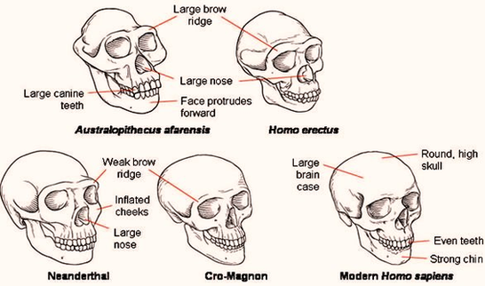
Skull Comparisons refers to a series of image macros that parody diagrams that compare skulls of the different stages of human evolution.

The twist is that they all look the same except one. Add your less evolved skull description to complete the meme. Click above to edit this template directly in your browser. Easily remix this template with your own text, images, and videos.

The final cranial bone is the sphenoid bone shown in light purple/pink below. It is the only cranial bone that articulates with all of the other cranial bones. The sphenoid bone is difficult to see on the side view of the skull, but it is actually shaped like a butterfly Let’s look at other slices of the skull to better understand the shape.

Diagram Skull Comparison Image Diagram - Chart - diagrams and charts with labels. This diagram depicts Diagram Skull Comparison Image

Diagram Skull Comparison Image

Picture Of The Skeletal System

Picture Of The Skeletal System Diagram - Chart - diagrams and charts with labels. This diagram depicts Picture Of The Skeletal System

picture of the skeletal system

Skeleton Names

Best Skeleton Names The Big Le-bone-ski Bones McCoy Teddy Bones-evelt Bury White Clarence Marrow Johnny Rotten Jon Bone Jovi Hugh Maris Indiana Bones Blake Skeleton Clay Vackle Albert Spinestein Vincent Van Bone Scary Potter Jim Reaper Eddie Deader Doug Upp Nefarious Bueller Werner Bray Bone-jangles …

One of the (plastic cast) skeletons in my classroom is nicknamed Slim, and the other is Leonard (after the character Dr. Leonard H. “Bones” McCoy from the TV show Star Trek). The chimpanzee skeleton is “Bobo,” and the gorilla skeleton is “Magilla.”

Skeletons also provide ample opportunities for puns – make no bones about it. If you’re trying to figure out what to name your skeleton, you can find lots of inspiration in anatomical terms and corny Halloween wordplay. Or, better yet, you can just search the following list of funny skeleton names for the perfect idea.

Skeleton Names Diagram - Chart - diagrams and charts with labels. This diagram depicts Skeleton Names

skeleton names

Skeletal Muscles Diagram

Attached to the bones of the skeletal system are about 700 named muscles that make up roughly half of a person’s body weight. Each of these muscles is a discrete organ constructed of skeletal muscle tissue, blood vessels, tendons, and nerves.

This is a table of skeletal muscles of the human anatomy. There are around 640 skeletal muscles within the typical human body.

Cardiac and skeletal muscles are “striated” in that they contain sarcomeres and are packed into highly regular arrangements of bundles; smooth muscle has neither.

Skeletal Muscles Diagram Diagram - Chart - diagrams and charts with labels. This diagram depicts Skeletal Muscles Diagram

skeletal muscles diagram

Skeletal Muscle Chart

skeletal muscle, which exists throughout the body The body’s collective muscle tissue constitutes its muscle mass. However, in most contexts, the term “muscle mass” specifically refers to skeletal muscle. This is the only type of muscle that a person can voluntarily control.

Skeletal muscle is a fascinating tissue with a complex structure. It consists of elongated multinuclear cells called the myocytes (or myofibers). The muscle cells can be anything from 1 mm to 30 cm in length. The longest muscle cell in our bodies can be found in the sartorius muscle and is 30 cm (nearly 12 inches!) long. From biology-forums.com

There are around 640 skeletal muscles within the typical human body. Almost every muscle constitutes one part of a pair of identical bilateral muscles, found on both sides, resulting in approximately 320 pairs of muscles, as presented in this article.

Skeletal Muscle Chart Diagram - Chart - diagrams and charts with labels. This diagram depicts Skeletal Muscle Chart

skeletal muscle chart

Unlabeled Human Skeleton

Labeled Human Skeleton. The labeled human skeleton system is comprised of 206 different bones of various sizes and shapes, all with the primary purpose of providing support, protection, and shape to the human body. When humans are born we have close to 300 bones, and over time they fuse together.

Written By: Human skeleton, the internal skeleton that serves as a framework for the body. This framework consists of many individual bones and cartilages. There also are bands of fibrous connective tissue—the ligaments and the tendons—in intimate relationship with the parts of the skeleton.

The labeled human skeleton system is comprised of 206 different bones of various sizes and shapes, all with the primary purpose of providing support, protection, and shape to the human body. When humans are born we have close to 300 bones, and over time they fuse together.

Unlabeled Human Skeleton Diagram - Chart - diagrams and charts with labels. This diagram depicts Unlabeled Human Skeleton

unlabeled human skeleton

Skull Diagram Anatomy

Labeled Skull Diagram. The idea behind using labeled diagrams is to get an overview of all of the structures within a given area. When it comes to testing your memory of these structures, previously having seen them altogether as a group should help you to remember them more easily.

Human Organ Systems Diagram – Human Organ Systems Chart – Human anatomy diagrams and charts explained. This diagram depicts Human Organ Systems with parts and labels. Human Muscles Diagram – Human Muscles Chart – Human anatomy diagrams and charts explained. This diagram depicts Human Muscles with parts and labels.

These are paired bones, with the right and left parietal bones joining together at the top of the skull. Each parietal bone is also bounded anteriorly by the frontal bone, inferiorly by the temporal bone, and posteriorly by the occipital bone. Temporal Bone Figure 5. Temporal Bone.

Skull Diagram Anatomy Diagram - Chart - diagrams and charts with labels. This diagram depicts Skull Diagram Anatomy

skull diagram anatomy

Pics Of Skeletal System

Regardless of age or sex, the skeletal system can be broken down into two parts, known as the axial skeleton and the appendicular skeleton. The adult axial skeleton consists of 80 bones. It’s made up of the bones that form the vertical axis of the body, such as the bones of the head, neck, chest, and spine. The adult skull comprises 22 bones.

The skeleton acts as a scaffold by providing support and protection for the soft tissues that make up the rest of the body. The skeletal system also provides attachment points for muscles to allow movements at the joints.

Providing Physical Structure And Canine Skeletal System X-ray, lungs, skeletal system,. The marked region on the image display is made to increase the effectiveness it is not marked by a physician Skeleton with muscles side.

Pics Of Skeletal System Diagram - Chart - diagrams and charts with labels. This diagram depicts Pics Of Skeletal System

pics of skeletal system

Skeleton And Organs Diagram

Written By: Human skeleton, the internal skeleton that serves as a framework for the body. This framework consists of many individual bones and cartilages. There also are bands of fibrous connective tissue—the ligaments and the tendons—in intimate relationship with the parts of the skeleton.

The motion is limited by the ligamentous attachments between ribs and vertebrae. The third general function of the skeleton is that of motion. The great majority of the skeletal muscles are firmly anchored to the skeleton, usually to at least two bones and in some cases to many bones.

The Human Body 1 TRACHEA. 2 LUNGS. 3 HEART. 4 LIVER. 5 STOMACH. 6 SPLEEN. 7 GALLBLADDER. 8 KIDNEYS. 9 BLADDER. 10 SMALL INTESTINES. More items…

Skeleton And Organs Diagram Diagram - Chart - diagrams and charts with labels. This diagram depicts Skeleton And Organs Diagram

skeleton and organs diagram

Skeleton Human

Author of Textbook of Comparative Histology. Human skeleton, the internal skeleton that serves as a framework for the body. This framework consists of many individual bones and cartilages. There also are bands of fibrous connective tissue —the ligaments and the tendons —in intimate relationship with the parts of the skeleton.

The Adult Human Skeleton Is Made Up of 206 Bones. These bones provide structure and protection and facilitate motion. Bones articulate to form structures. The skull protects the brain and gives shape to the face.

Each bone is a complex living organ that is made up of many cells, protein fibers, and minerals. The skeleton acts as a scaffold by providing support and protection for the soft tissues that make up the rest of the body. The skeletal system also provides attachment points for muscles to allow movements at the joints.

Skeleton Human Diagram - Chart - diagrams and charts with labels. This diagram depicts Skeleton Human

skeleton human

What Are The Functions Of The Skeletal System

The skeletal system is the body system composed of bones and cartilage and performs the following critical functions for the human body: supports the body. facilitates movement. protects internal organs. produces blood cells. stores and releases minerals and fat.

The skeletal system’s main function is to provide support for the body. For example, the spinal column provides support for the head and torso. The legs, on the other hand, support and bear the weight of the upper body while a person stands.

The muscular and skeletal systems work together as the musculoskeletal system, which enables body movement and stability. When muscles contract, they pull on bones of the skeleton to produce movement or hold the bones in a stable position.

What Are The Functions Of The Skeletal System Diagram - Chart - diagrams and charts with labels. This diagram depicts What Are The Functions Of The Skeletal System

what are the functions of the skeletal system

Skeleton Chart

The Human Skeletal System Chart shows anterior, lateral and posterior views of the skeletal system.

This printout will help you learn about the human skeleton and identify some of the important features of our skeletal anatomy. This document contains an outline of a juvenile human standing 109 cm tall (or 43” tall). Modern humans average a height of 109 cm between 5 and 6 years of age.

The motion is limited by the ligamentous attachments between ribs and vertebrae. The third general function of the skeleton is that of motion. The great majority of the skeletal muscles are firmly anchored to the skeleton, usually to at least two bones and in some cases to many bones.

Skeleton Chart Diagram - Chart - diagrams and charts with labels. This diagram depicts Skeleton Chart

skeleton chart

Skeleton Labeled Diagram

Labeled Human Skeleton. The labeled human skeleton system is comprised of 206 different bones of various sizes and shapes, all with the primary purpose of providing support, protection, and shape to the human body. When humans are born we have close to 300 bones, and over time they fuse together.

Given below is a labeled diagram, and tips to help you draw and memorize the names of different parts. Here is a detailed diagram which shows the various bones present in an adult skeletal system. There is a little difference between the male and female skeleton, but for diagrams mostly a male skeletal system is considered.

The skeletal system is one of the important human body systems. We have divided the human skeletal system, into four sections for better understanding, so let us go from top to bottom and study the human skeleton.

Skeleton Labeled Diagram Diagram - Chart - diagrams and charts with labels. This diagram depicts Skeleton Labeled Diagram

skeleton labeled diagram

Skeletal Pictures

Teeth are not bones but are still part of the human skeletal system. While the adult human skeletal system includes 206 bones, the infantile skeletal system has many more, since not all of the bones have fused together yet.

The skeleton acts as a scaffold by providing support and protection for the soft tissues that make up the rest of the body. The skeletal system also provides attachment points for muscles to allow movements at the joints.

Skeletal System 1 Skull. 2 Hyoid and Auditory Ossicles. 3 Vertebrae. 4 Ribs and Sternum. 5 Pectoral Girdle and Upper Limb. 6 Pelvic Girdle and Lower Limb. 7 Microscopic Structure of Bones. 8 Types of Bones. 9 Parts of Bones. 10 Articulations.

Skeletal Pictures Diagram - Chart - diagrams and charts with labels. This diagram depicts Skeletal Pictures

skeletal pictures

Picture Of Skull Bones

The bones that make up the cranium are called the cranial bones. The remainder of the bones in the skull are the facial bones. Figure 6.7 and Figure 6.8 show all the bones of the skull, as they appear from the outside. In Figure 6.9, some of the bones of the hard palate forming the roof of the mouth are visible because the mandible is not present.

Occipital bone: the bone that forms the back of the head and connects with the occipital condyles and foramen magnum — skeletal structures located on the underside of the skull, near the spine — and the lambdodial suture, which is at the back of the skull.

A collection of 22 bones, the skull protects the all-important brain and supports the other soft tissues of the head. During fetal development, the bones of the skull form within tough, fibrous membranes in a fetus’ head. As these bones grow throughout fetal and childhood development, they begin to fuse together, forming a single skull.

Picture Of Skull Bones Diagram - Chart - diagrams and charts with labels. This diagram depicts Picture Of Skull Bones

picture of skull bones

Skeletal Muscle Picture 2

Skeletal muscle, also called voluntary muscle, in vertebrates, most common of the three types of muscle in the body. Skeletal muscles are attached to bones by tendons, and they produce all the movements of body parts in relation to each other. Unlike smooth muscle and cardiac muscle, skeletal muscle is under voluntary control.

Skeletal muscle, also called voluntary muscle, in vertebrates, most common of the three types of muscle in the body.

[edit on Wikidata] Skeletal muscle is one of three major muscle types, the others being cardiac muscle and smooth muscle. It is a form of striated muscle tissue, which is under the voluntary control of the somatic nervous system. Most skeletal muscles are attached to bones by bundles of collagen fibers known as tendons.

Skeletal Muscle Picture 2 Diagram - Chart - diagrams and charts with labels. This diagram depicts Skeletal Muscle Picture 2

skeletal muscle picture 2

Pictures Of Skeletal System

Skeletal System 1 Skull. 2 Hyoid and Auditory Ossicles. 3 Vertebrae. 4 Ribs and Sternum. 5 Pectoral Girdle and Upper Limb. 6 Pelvic Girdle and Lower Limb. 7 Microscopic Structure of Bones. 8 Types of Bones. 9 Parts of Bones. 10 Articulations.

The bones of the skeletal system act as attachment points for the skeletal muscles of the body. Almost every skeletal muscle works by pulling two or more bones either closer together or further apart. Joints act as pivot points for the movement of the bones.

The skeleton acts as a scaffold by providing support and protection for the soft tissues that make up the rest of the body. The skeletal system also provides attachment points for muscles to allow movements at the joints. New blood cells are produced by the red bone marrow inside of our bones.

Pictures Of Skeletal System Diagram - Chart - diagrams and charts with labels. This diagram depicts Pictures Of Skeletal System

pictures of skeletal system

Skeleton Foot

The skeleton of the foot from behind forward is composed of the following bones: Tarsals. Metatarsals. Phalanges.

Identification of bones in the skeleton of the foot: Calcaneus (heel bone) is the largest and most proximal bone. Talus is the 2nd largest bone and is located above the calcaneus like a rider, therefore maximum bone in the skeleton of foot. Navicular is boat-shaped and is located in front of the head of talus. Cuboid is cubical in…

Easy Notes On 【Skeleton of the Foot】Learn in Just 6 Minutes! – Earth’s Lab The skeleton of the foot from behind forward is composed of the following bones: Tarsals. Metatarsals. Phalanges. All these are short bones which collectively create tarsus. These arearranged in 3 rows: (a) Proximal row includes talus and calcaneus.

Skeleton Foot Diagram - Chart - diagrams and charts with labels. This diagram depicts Skeleton Foot

skeleton foot