Human Eye Anatomy Orbit Pic Image

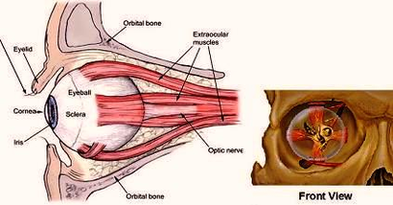
5,297 human eye anatomy 3d stock photos, vectors, and illustrations are available royalty-free. See human eye anatomy 3d stock video clips

In anatomy, the orbit is the cavity or socket of the skull in which the eye and its appendages are situated. “Orbit” can refer to the bony socket, or it can also be used to imply the contents.

Vector illustration. human eye anatomy stock illustrations 3d illustration of eye anatomy The human eye is an organ which reacts to light and pressure. As a sense organ, the mammalian eye allows vision. Human eyes help provide a three dimensional, moving image, normally coloured in daylight.

Human Eye Anatomy Orbit Pic Image Diagram - Chart - diagrams and charts with labels. This diagram depicts Human Eye Anatomy Orbit Pic Image

Human Eye Anatomy Orbit Pic Image

Week Pregnancy Pic Image

You can clearly see two little feet in this scan picture, fully formed at 21 weeks. The baby’s facial profile can be clearly seen in this image. In this 3D image of a 23-week old fetus, the baby seems to be yawning! You can also buy special photo albums to hold your baby’s scan pictures.

The fetus here is pictured with the head lowered towards the chest. At 10 weeks, the fetus measures approximately 2.5 centimetres. The head is still huge in size compared to the rest of the body. In this picture, the head is still inclined towards the chest, but soon it’ll gradually begin to raise.

A baby in the first trimester at 12 weeks has the following features: Muscles and nerves are coordinated. Baby can make a fist. The baby’s eyelids are closed and will not open until 28 weeks’ gestation. The growth of the head is slower and the baby has grown longer. Your baby weighs nearly one ounce and is about 3 inches long.

Week Pregnancy Pic Image Diagram - Chart - diagrams and charts with labels. This diagram depicts Week Pregnancy Pic Image

Week Pregnancy Pic Image

Xray Normal Ankle Pic Image

Ankle x-ray. Close-up of a ankle x-ray Human foot ankle and leg in x-ray, on gray background. The foot ankle is highlighted by green colour X-ray ankle show fracture distal tibia and fibula (leg’s bone).

Human foot ankle and leg in x-ray, on gray background. The foot ankle is highlighted by green colour X-ray ankle show fracture distal tibia and fibula (leg’s bone). Film x-ray ankle show fracture distal tibia and fibula (leg’s bone

Lateral view of the ankle tracing the outline of the tibia (green), fibula (red), talus (dark red), calcaneum (blue) and base of 5th metatarsal (orange). Note the os trigonum (red circle) which is an accessory ossicle of the foot and a normal variant. 3 Assess the joint space on the mortise view.

Xray Normal Ankle Pic Image Diagram - Chart - diagrams and charts with labels. This diagram depicts Xray Normal Ankle Pic Image

Xray Normal Ankle Pic Image