Diagram Floating Shoulder Xray Image

This human anatomy diagram with labels depicts and explains the details and or parts of the Diagram Floating Shoulder Xray Image. Human anatomy diagrams and charts show internal organs, body systems, cells, conditions, sickness and symptoms information and/or tips to ensure one lives in good health.

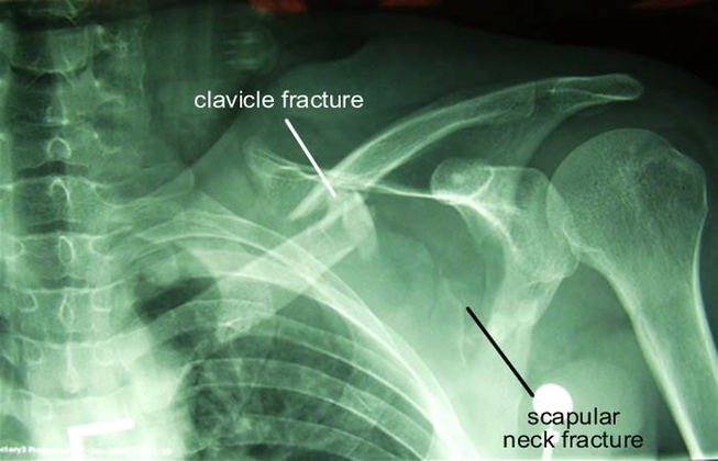
Typical shoulder X-ray views include: An axial view can also be used as an alternative to the scapula Y view if the patient is unable to tolerate the positioning required to obtain this view. Figure 1. A normal AP view 1 Figure 1.1.

Typical X-ray findings in posterior shoulder dislocation include: AP view: the glenohumeral joint will be widened and the humeral head will take on a classic “light bulb” appearance due to forced internal rotation of the humerus. Lateral view: the humeral head will lie posterior to the glenoid fossa.

The shoulder external rotation view is an additional projection to the standard shoulder series it is often combined with the internal rotation view to visualize the entirety of the humeral head. This projection best demonstrates the greater tubercle humerus in profile. Article:

Diagram Floating Shoulder Xray Image

Diagram Floating Shoulder Xray Image


Tags: , ,