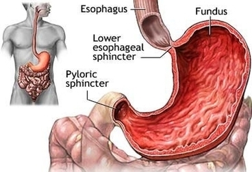
Stomach Lymphnodes Image
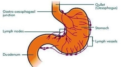
Gastric Lymph Nodes: There are numerous gastric lymph node groups. They drain the stomach, upper duodenum, abdominal oesophagus and the greater omentum into the coeliac group. Hepatic Lymph Nodes: The hepatic nodes extend in the lesser omentum along the hepatic View Diagram Stomach Lymphnodes Image