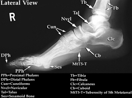
A normal foot x-ray will show three images of the foot, one from the front, one from the side, and one from the angle. The gamma ring is the smallest gamma ray and is not absorbed by all tissues. The gamma arcs cannot penetrate through dense structures, shielding the film from the rays.
The patient can remain supine with an image receptor placed vertically adjacent to the lateral aspect of the upright ankle, and the x-ray beam is directed horizontally, centered at the bony prominence of the medial malleolus of the distal tibia.
Age: 20 years. Loading images… Normal right foot radiographs in a young adult female for reference. Normal right foot radiographs in a young adult female for reference.
Foot Normal Lat Xray Image Diagram - Chart - diagrams and charts with labels. This diagram depicts Foot Normal Lat Xray Image
