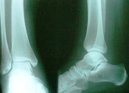
Ankle x-ray. Close-up of a ankle x-ray Human foot ankle and leg in x-ray, on gray background. The foot ankle is highlighted by green colour X-ray ankle show fracture distal tibia and fibula (leg’s bone). Xray Normal Ankle Pic Image Diagram - Chart - diagrams and charts with labels. This diagram depicts Xray Normal Ankle Pic Image and explains the details of Xray Normal Ankle Pic Image.
Xray Normal Ankle Pic Image