
Category Archives: Anatomy
Crown And Bridge1 Image
Anatomy And Physiology Of Animals Growing Bone Image
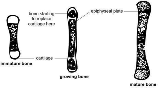
138Anatomy and physiology of domestic animals associated with bone formation, it can occur in other tissues. There are two general classes of bone formation. Intramembranous ossifi cation occurs when bone develops from a fi brous membrane. The fl at bones View Diagram Anatomy And Physiology Of Animals Growing Bone Image
Human Muscle Diagram Image
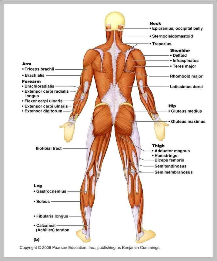
105,188 human muscle anatomy stock photos, vectors, and illustrations are available royalty-free. Anatomy front view of major face muscles of a woman including procerus, masseter, orbicularis oculi, zygomaticus, buccinator and nasalis. Leg, Hip & Gluteal Anatomy 1 Gluteal Muscles 2 View Diagram Human Muscle Diagram Image
Diagram Longbone Image
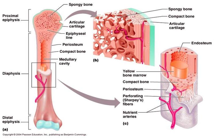
Image of a typical long bone is shown with numbers identifying the various parts, such as the epiphysis. Blank Skull Diagram – Blank Long Bone Diagram Popular Skull diagram unlabeled skull diagram inferior view template information title. First, what is View Diagram Diagram Longbone Image
Human Brain Development Timeline Image

The human brain at stages 18-20, including the choroid plexuses and the amygdaloid and septal nuclei. Anat. Embryol. , 182, 285-306. PMID: 2268071 â 25.0 25.1 Müller F & O’Rahilly R. (1990). The human brain at stages 21-23, with particular View Diagram Human Brain Development Timeline Image
Diabetes Glycemic Index Image

Glycemic index and diabetes. Glycemic index (GI) is a measure of how quickly a food can make your blood sugar (glucose) rise. Only foods that contain carbohydrates have a GI index. The GI values can be broken down into three View Diagram Diabetes Glycemic Index Image
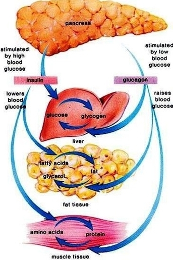
Diabetes Mellitus Insulin Treatment Image

InsulinInsulin Replacement Therapy Many people with diabetes require drugs to lower blood glucose levels, relieve symptoms, and prevent complications of diabetes. There are two types of diabetes mellitus Type 1, in which the… read more is the most commonly used View Diagram Diabetes Mellitus Insulin Treatment Image
Baby Development Week By Week Image
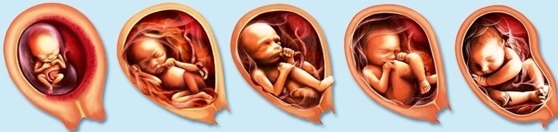
What You’re Seeing: This 3D image of your developing baby shows how lifelike she appears at this early age. Notice that baby-to-be is tucked into a c-shape, with her head toward her stomach and her arms and legs jutting outward. View Diagram Baby Development Week By Week Image
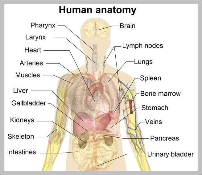
Show A Diagram Of The Human Body Image

43,556 human body diagram stock photos, vectors, and illustrations are available royalty-free. internal organs and circulatory system Vector isolated illustration of human internal organs and circulatory system in man body. Stomach, liver, bladder, lung, kidney, heart, icon. Medical poster organs View Diagram Show A Diagram Of The Human Body Image
Crukmig1 Image

Diabetes Insipidus Treatment Image
What is diabetes insipidus? Diabetes insipidus is a condition that results from insufficient production of the antidiuretic hormone (ADH), a hormone that helps the kidneys and body conserve the correct amount of water. Normally, the antidiuretic hormone controls the kidneys’ View Diagram Diabetes Insipidus Treatment Image
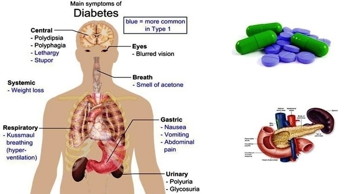
Type Diabetes Mellitus Symptoms Causes Image
Symptoms & Causes of Diabetes. What are the symptoms of diabetes? Symptoms of diabetes include. increased thirst and urination. increased hunger. fatigue. blurred vision. numbness or tingling in the feet or hands. Symptoms of type 2 diabetes often develop slowly—over View Diagram Type Diabetes Mellitus Symptoms Causes Image
Ear Anatomy1 Image
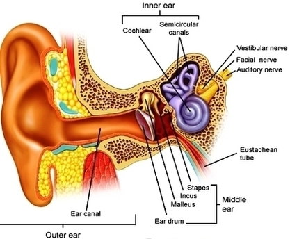
1,061 ear anatomy stock photos and images available, or search for anatomy model or muscle anatomy to find more great stock photos and pictures. Inner Ear Anatomy The inner ear is where the sound waves are translated into types of View Diagram Ear Anatomy1 Image
Anatomy Foot Ankle Image
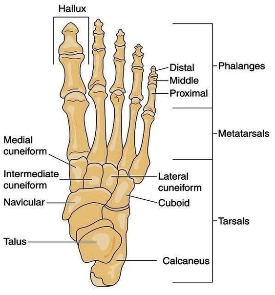
The anatomy of the foot is incredibly complex. This introduction to the anatomy of the foot and ankle will be very general and highlight the most relevant structures. The ankle joint or tibiotalar joint is formed where the top of View Diagram Anatomy Foot Ankle Image
Cochlear Duct Image
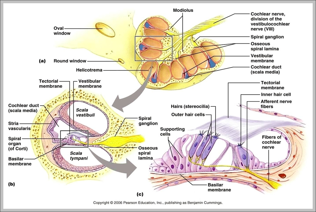
The Cochlear Duct (or Scala Media) is an endolymph filled cavity inside the cochlea, located in between the tympanic duct and the vestibular duct, separated by the basilar membrane and Reissner’s membrane (the vestibular membrane) respectively. The cochlear duct subdivides View Diagram Cochlear Duct Image
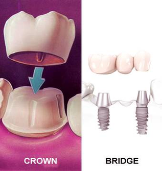
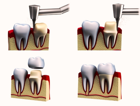
Dental Crowns Procedure Image
Bronchi Lungs Diagram Image
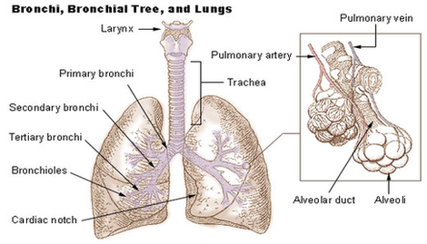
What are Bronchi. Bronchi, branching from the trachea, are the primary passageway for air to get into the lungs [1]. It is the plural for bronchus. Each bronchus further branches into smaller tubes or bronchioles. There are two primary (extrapulmonary) View Diagram Bronchi Lungs Diagram Image
B0001404 Five Week Old Human Embryo
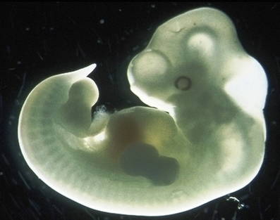
It measures 0.118 inches (3 mm) from crown to rump — basically, from head to bum. Your five-week embryo might be tiny, but there are many exciting things starting to take shape! Embryonic development in the human, covers the first View Diagram B0001404 Five Week Old Human Embryo
Diagram Of Human Anatomy Mediastinum Left View Illustration Large Image
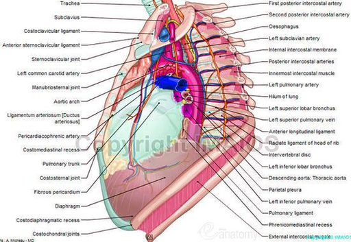
Nerves: details the nervous anatomy of the mediastinum including notably the route of the phrenic and vagus nerves as well as the anatomy of the sympathetic trunk at the thoracic level. Heart: the external morphology of the heart in the View Diagram Diagram Of Human Anatomy Mediastinum Left View Illustration Large Image