Major Respiratory Organs
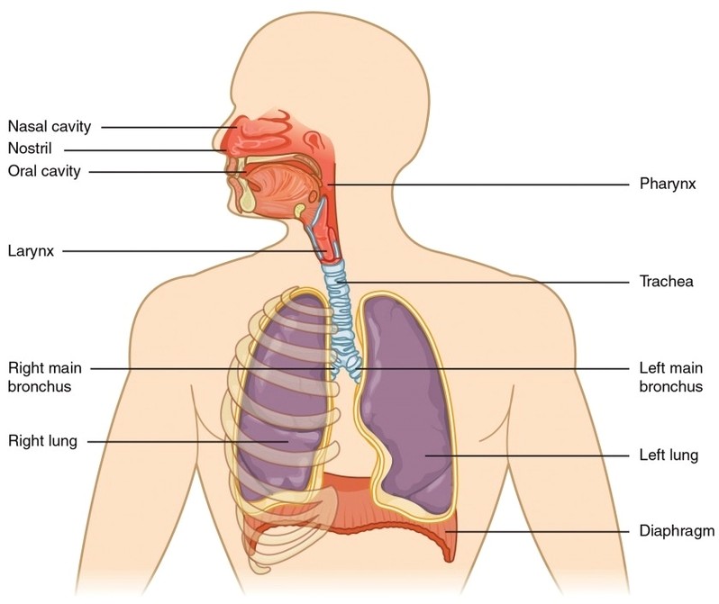
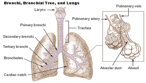
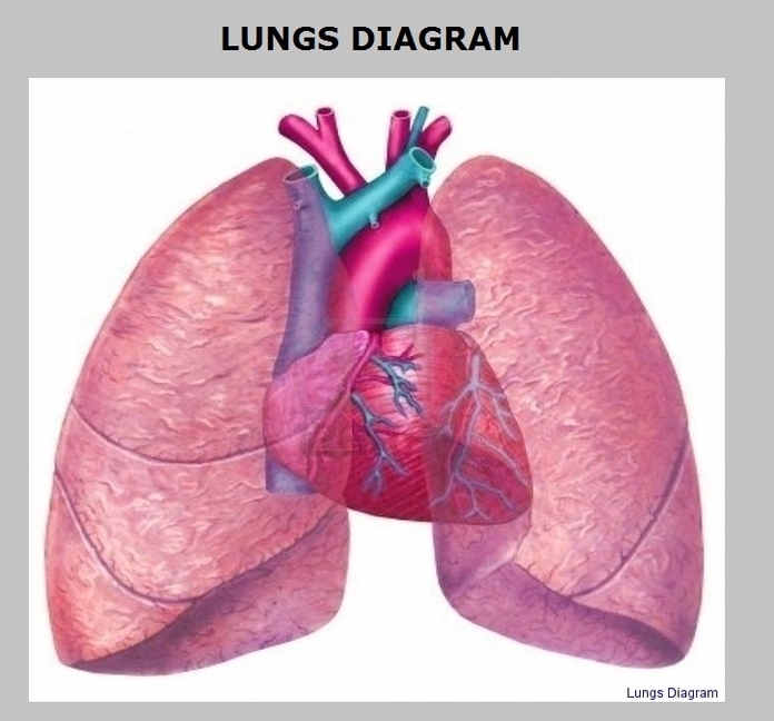
Major Respiratory Organs: Major respiratory organs include the nose, pharynx, larynx, trachea, bronchi, and lungs, all working together to move air, filter particles, and facilitate gas exchange.