1,061 ear anatomy stock photos and images available, or search for anatomy model or muscle anatomy to find more great stock photos and pictures.
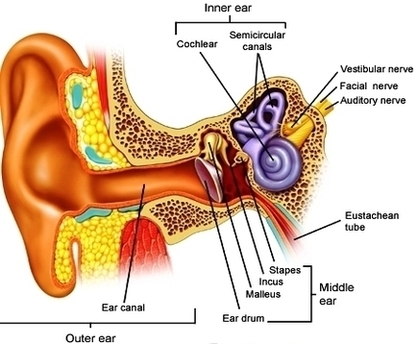
Inner Ear Anatomy The inner ear is where the sound waves are translated into types of electrical nerve impulses. Most of the hearing and balance content is located within the bony labyrinth. After the tympanic membrane, these are the nerves that most likely contribute to hearing impairment and may require treatment or medical services.
The ear has external, middle, and inner portions. The outer ear is called the pinna and is made of ridged cartilage covered by skin. Sound funnels through the pinna into the external auditory canal, a short tube that ends at the eardrum (tympanic membrane).
Ear Anatomy1 Image

