Cochlear Duct Image

This human anatomy diagram with labels depicts and explains the details and or parts of the Cochlear Duct Image. Human anatomy diagrams and charts show internal organs, body systems, cells, conditions, sickness and symptoms information and/or tips to ensure one lives in good health.

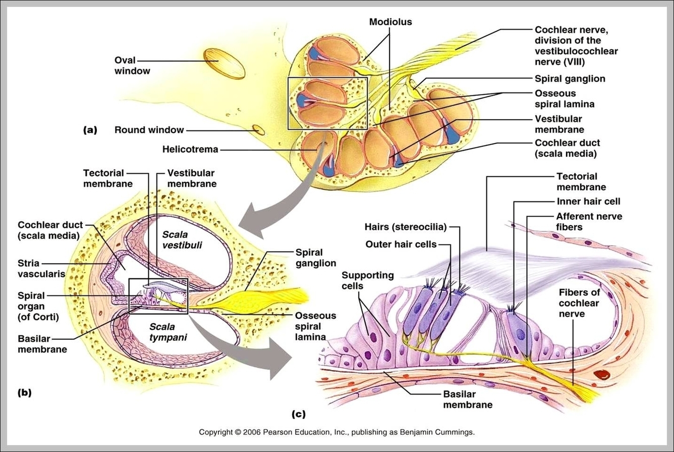
The Cochlear Duct (or Scala Media) is an endolymph filled cavity inside the cochlea, located in between the tympanic duct and the vestibular duct, separated by the basilar membrane and Reissner’s membrane (the vestibular membrane) respectively.

The cochlear duct subdivides the bony labyrinth into two perilymph chambers, namely the scala vestibuli anteriorly (opens into the vestibule) and the scala tympani posteriorly (ends at the round window) 1. It is separated from the scala vestibuli by Reissner’s membrane (vestibular membrane) 5 .

In previous studies, it was common to express the diameter of the cochlea in measurements along two, four, or eight radii 5, 6, 17. Using 721 assessments, one per angular angle, the tapering course of the cochlear walls can be mapped more accurately.

Cochlear Duct Image

Cochlear Duct Image


Tags: ,