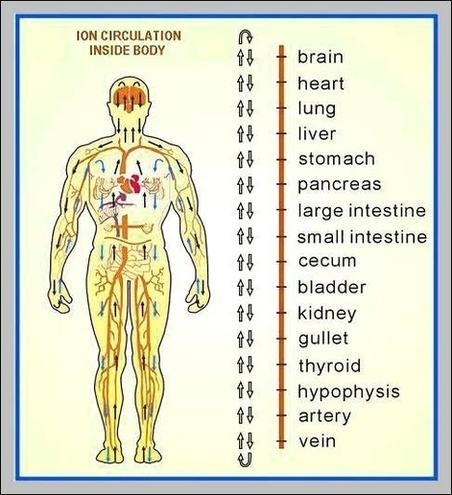
Human Body Layout Image
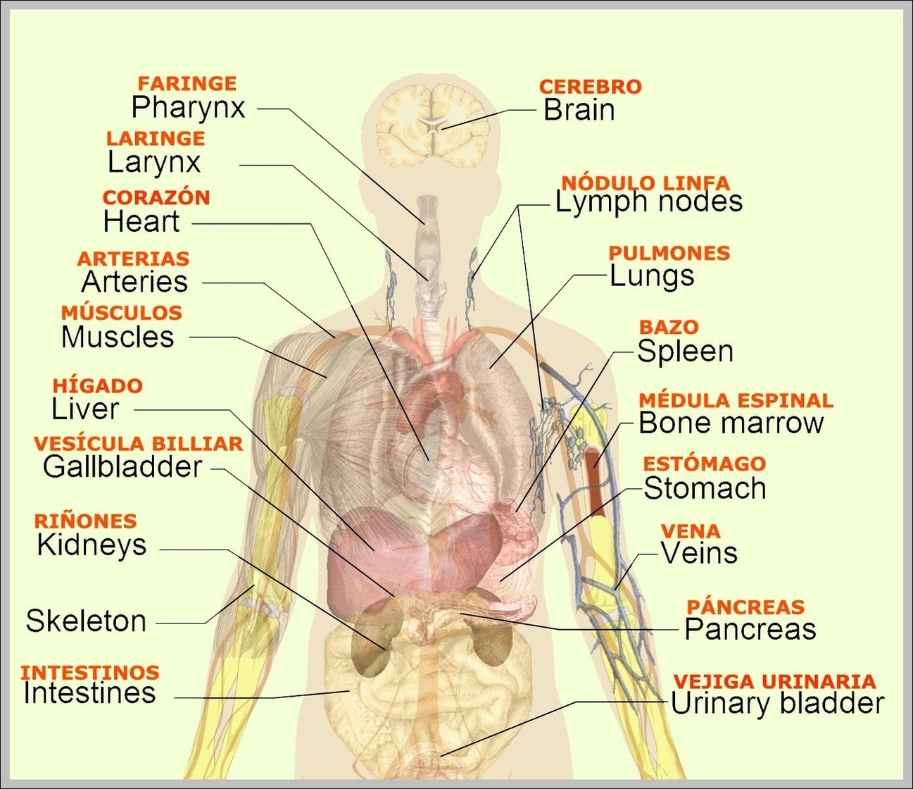
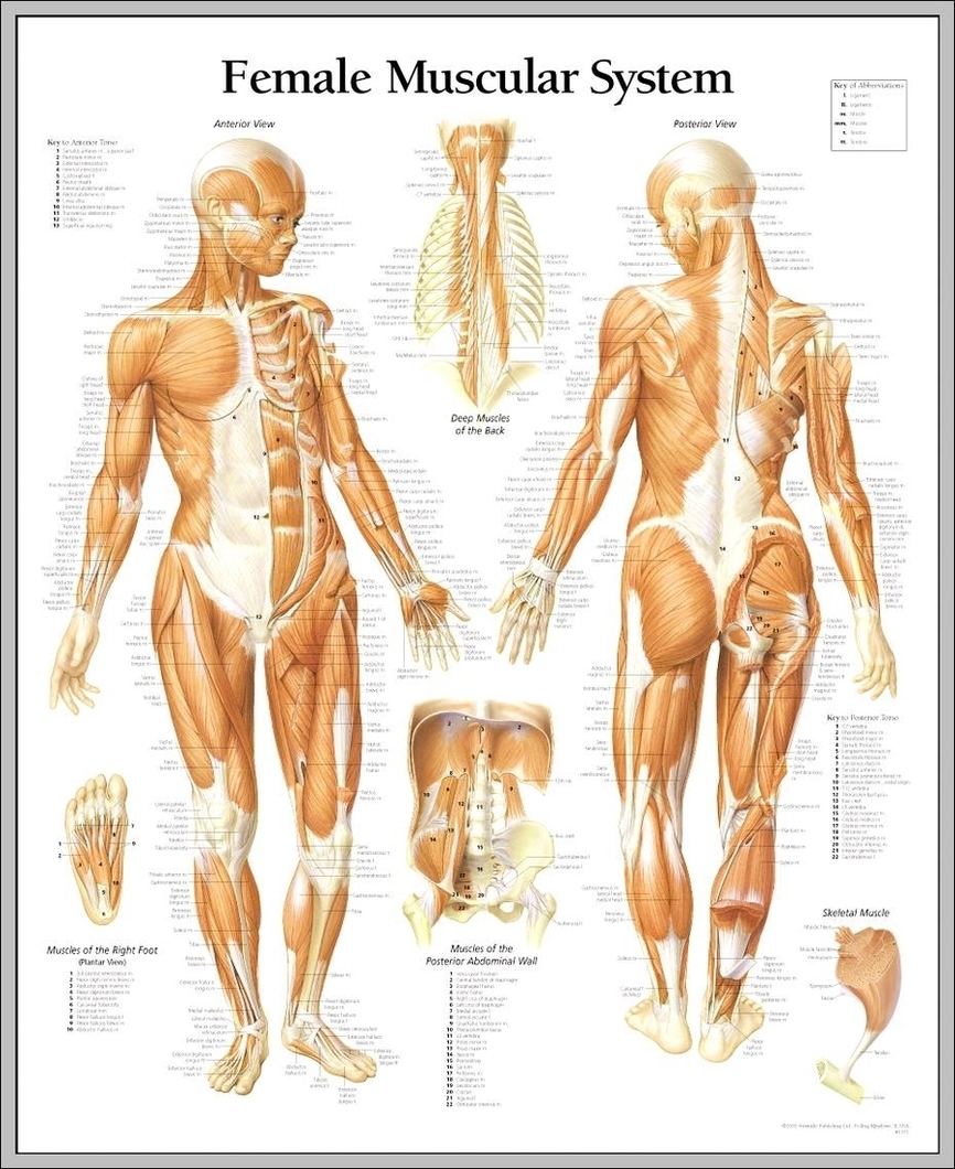
43,556 human body diagram stock photos, vectors, and illustrations are available royalty-free. 36,060 pictures of the human body stock photos, vectors, and illustrations are available royalty-free. Designed for easy recognition of functional aspects of the human body, the labeled human View Diagram Human Body Layout Image