Category Archives: Anatomy
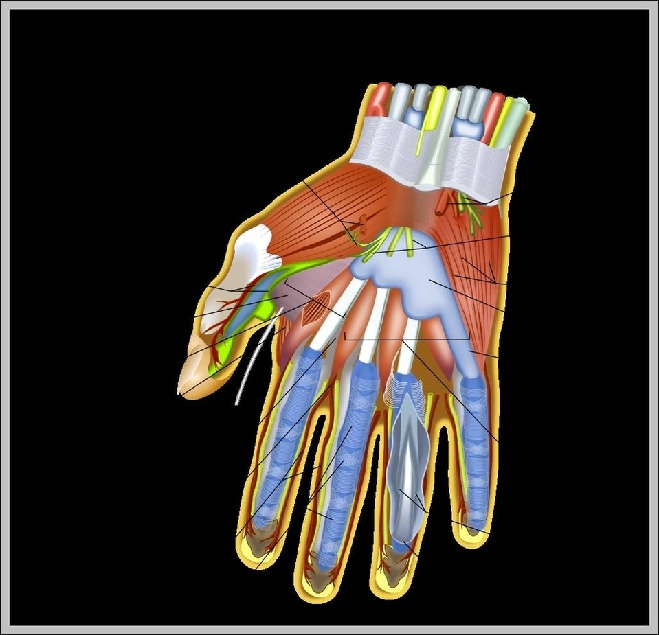
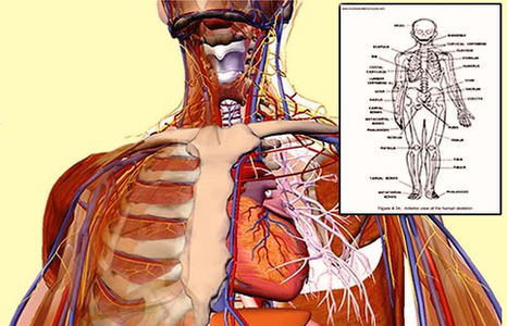
Human Anatomy Tendons And Ligaments Image
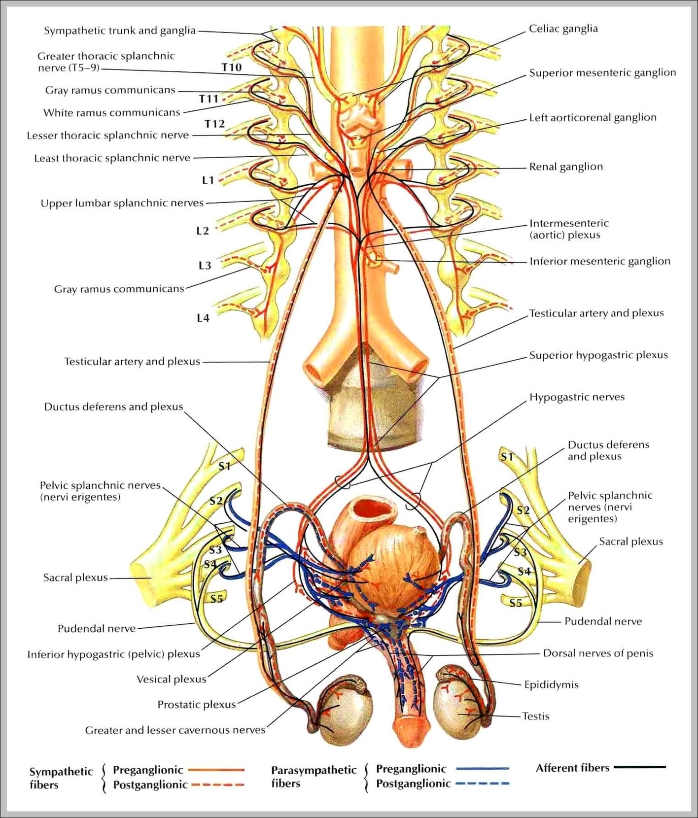
Male Organs Anatomy Image
Brainor Webt Image
Positive Pregnancy Test Image
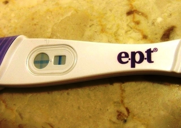
17,643 positive pregnancy test stock photos, vectors, and illustrations are available royalty-free. See positive pregnancy test stock video clips Here Are 12 Pictures of Positive Pregnancy Tests to Compare Yours To If you take a pregnancy test, especially if you View Diagram Positive Pregnancy Test Image
Human Muscles Image

93,451 human body muscles stock photos and images available, or search for anatomy or human anatomy to find more great stock photos and pictures. Last Updated: Jul 16, 2019 The muscular system is responsible for the movement of the human View Diagram Human Muscles Image
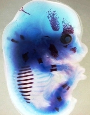
Species Fetus Image
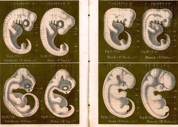
10,383 fetus stock photos and images available or search for fetus icon or embryo to find more great stock photos and pictures. Wikimedia Commons has media related to Fetus. Prenatal Image Gallery Index at the Endowment for Human Development website, View Diagram Species Fetus Image
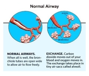
Stock Lungexarion Image
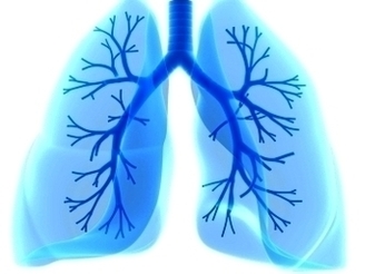
59,106 human lung stock photos and images available, or search for human lung anatomy or human lung illustration to find more great stock photos and pictures. 3D illustration of Lungs, medical concept. through our collection of LUNG pictures. All our View Diagram Stock Lungexarion Image
Plantar Fasciitis Pain Map Tt Tennis Warehouse Com Image
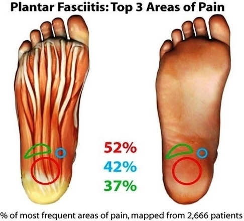
1 Overview. Plantar fasciitis is an inflammation of the fibrous tissue (plantar fascia) along the bottom of your foot that connects your heel bone to your toes. 2 Symptoms. Plantar fasciitis typically causes a stabbing pain in the bottom of View Diagram Plantar Fasciitis Pain Map Tt Tennis Warehouse Com Image
Ohmes Tooth Prepared For Crown Image
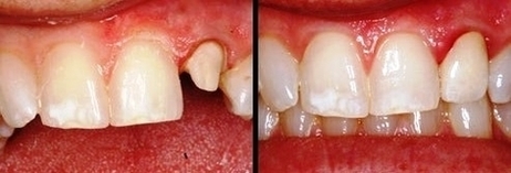
How is Your Tooth Prepared for a Dental Crown? Restoring one or several teeth to good form and function requires, besides other procedures the preparation of the teeth for placement of the indirect dental restorations (crown or bridge). This dental View Diagram Ohmes Tooth Prepared For Crown Image
Sciatic Nerve Picture Image
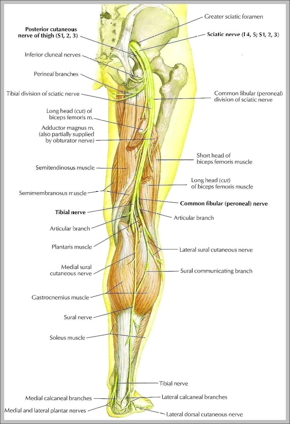
1,041 sciatic nerve stock photos and images available, or search for sciatic nerve pain or sciatic nerve diagram to find more great stock photos and pictures. piriformis syndrome 3d medical vector illustration on white… piriformis syndrome 3d medical vector illustration View Diagram Sciatic Nerve Picture Image
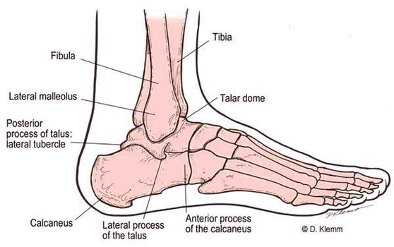
Diagram Lateral Foot Anatomy Image
Free Human Anatomy Diagrams Miewiiy Image
Te Nose Diagram Image
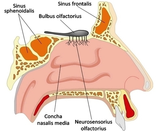
External Structure The surface of the human nose consists of a frontal portion comprised of the glabella, nasion, alar sidewalls and tip points; a basal portion made up of the columella, nostrils, soft tissues and infra tip lobule; and two View Diagram Te Nose Diagram Image
Diabetes Aids Carousel Image

Con: Carousels stand out from organic posts and are more obviously ads. On Facebook, multiple image posts take the form of a collage, with images arrayed in tiles. One image takes the stage, and the others wait in the wings View Diagram Diabetes Aids Carousel Image
Foot Bones Xray Ed Image
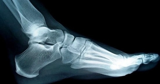
This x-ray is also beneficial for determining bone growth in children. The X-ray will help determine the size of the bones in the foot, as well as the size and position of bones and soft tissues in the foot. A View Diagram Foot Bones Xray Ed Image
Tooth Anatomy Page Image
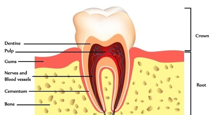
WebMD’s Teeth Anatomy Page provides a detailed diagram and definition of the teeth, inlcuding types, names, and parts of the teeth. Skip to main content Bodytomy provides labeled human tooth diagrams to help you understand the human tooth anatomy. The View Diagram Tooth Anatomy Page Image
Stomach Anatomy Ae Ba Ecfdalarge Image
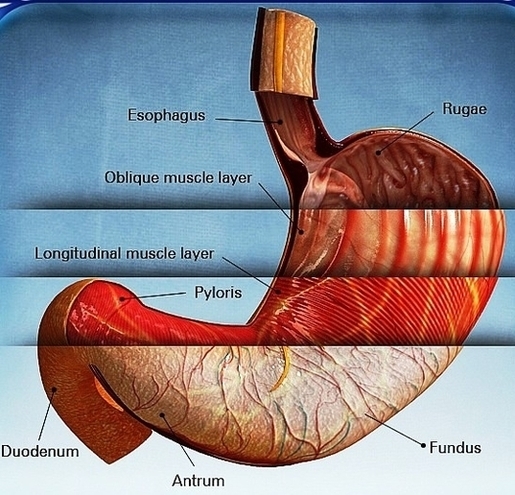
Diagram Of The Stages Of Pregnancy Month By Month Image
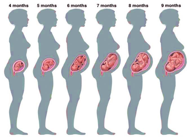
A typical pregnancy lasts 40 weeks from the first day of your last menstrual period (LMP) to the birth of the baby. It is divided into three stages, called trimesters: first trimester, second trimester, and third trimester. The fetus undergoes View Diagram Diagram Of The Stages Of Pregnancy Month By Month Image
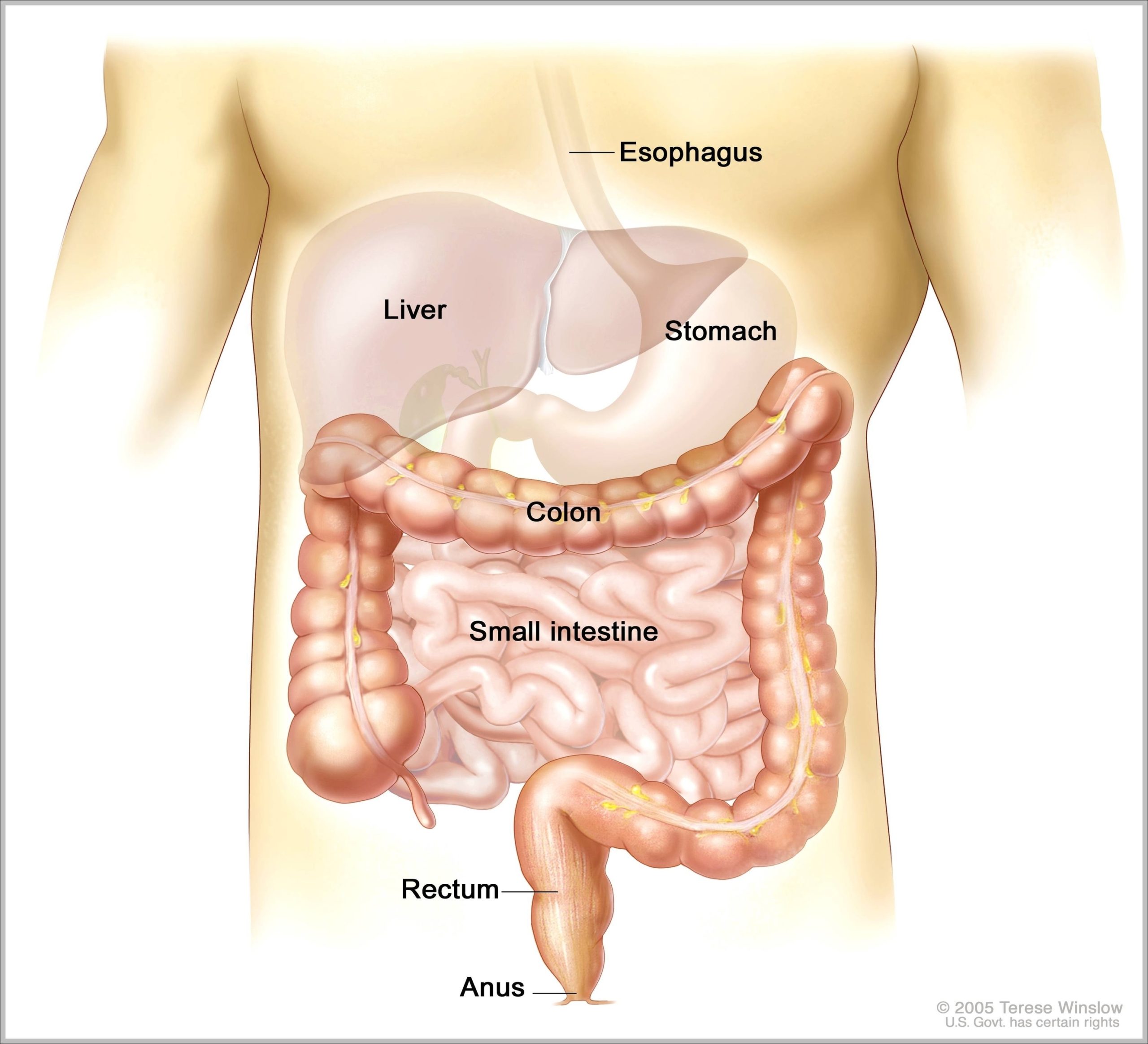
Picture Of Intestines Image