F Ear Anatomy Image

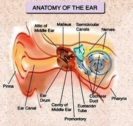
1,061 ear anatomy stock photos and images available, or search for anatomy model or muscle anatomy to find more great stock photos and pictures.

Picture of the Ear. The spiral-shaped cochlea is part of the inner ear; it transforms sound into nerve impulses that travel to the brain. The fluid-filled semicircular canals (labyrinth) attach to the cochlea and nerves in the inner ear. They send information on balance and head position to the brain.

Picture of the Ear. The fluid-filled semicircular canals (labyrinth) attach to the cochlea and nerves in the inner ear. They send information on balance and head position to the brain. The eustachian (auditory) tube drains fluid from the middle ear into the throat (pharynx) behind the nose.

F Ear Anatomy Image Diagram - Chart - diagrams and charts with labels. This diagram depicts F Ear Anatomy Image

F Ear Anatomy Image