Skip to content

Human Anatomy Diagrams

Menu

Primary menu

  • Top
  • Anatomy
  • Muscles
  • Systems
  • Body Parts
  • Organs
  • Cells
  • Bones
  • Teeth
  • Cancer
  • Health

Author Archives: anatomy

Early Forelimb Skeleton And Flesh Image

Posted on June 21, 2022 by anatomy
Early Forelimb Skeleton And Flesh Image

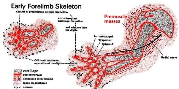
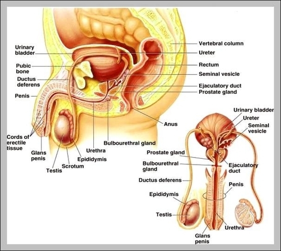
Anatomical landmarks: particular homologous structures on the skeleton (openings, joints, etc.) used for identifying the position of bones or other features of the anatomy. The skeleton of a dinosaur (or other vertebrate) is divided into a couple of different sections: View Diagram Early Forelimb Skeleton And Flesh Image→

Posted in Anatomy | Tagged early, flesh, forelimb, skeleton

Pictures Of The Male Anatomy Image

Posted on June 21, 2022 by anatomy

Pictures Of The Male Anatomy Image

Posted in Anatomy | Tagged male, pictures

Diagram Bones Of Foot Image

Posted on June 21, 2022 by anatomy
Diagram Bones Of Foot Image

When one looks at the anatomy of the foot, they would realize that the foot has a complex mechanical and structural architecture. The ankle joint is the shock absorber of the foot. Apart from 28 bones, 33 joints, muscles, ligaments, View Diagram Diagram Bones Of Foot Image→

Posted in Anatomy, Bones | Tagged bones, foot

Dental Implant Colgate Diagram Image

Posted on June 21, 2022 by anatomy
Dental Implant Colgate Diagram Image

The process requires the jawbone to heal tightly around the dental implant area (called osseointegration), absorbing it into the gumline as if it were a natural tooth. The implants are surgically placed within the jawbone and serve as the “root” View Diagram Dental Implant Colgate Diagram Image→

Posted in Anatomy | Tagged colgate, dental, implant

Blood Diagram Image

Posted on June 21, 2022 by anatomy

Blood Diagram Image

Posted in Anatomy | Tagged blood

Tooth Decay Photo Image

Posted on June 21, 2022 by anatomy

Tooth Decay Photo Image

Posted in Anatomy | Tagged decay, photo, tooth

Foot Plantar Fasciitis Causes Image

Posted on June 21, 2022 by anatomy
Foot Plantar Fasciitis Causes Image

Plantar fasciitis will usually start with a sudden onset of pain in the heel of your foot or under the ball of your foot. The most common causes of plantar fasciitis are: Those who carry extra body weight may be View Diagram Foot Plantar Fasciitis Causes Image→

Posted in Anatomy | Tagged Causes, fasciitis, foot, plantar

Testicles Pictures Image

Posted on June 21, 2022 by anatomy

Testicles Pictures Image

Posted in Anatomy | Tagged pictures, testicles

Real Pictures Of Human Organs Image

Posted on June 21, 2022 by anatomy

Real Pictures Of Human Organs Image

Posted in Anatomy, Organs | Tagged pictures, real

Eye section view

Posted on June 21, 2022 by anatomy
Eye section view

Eye – Cross Section. A human eyeball cross section showing the following structures: iris, anterior limiting membrane, posterior limiting membrane, tendon of Eye anatomy, Human eye cross section physiology, Model of cornea and lens for ophthalmologist. Eye anatomy, Human eye View Diagram Eye section view→

Posted in Anatomy, Organs | Tagged eye anatomy, eye diagram, Eye section view, human eye

The Musculoskeletal System Image

Posted on June 21, 2022 by anatomy
The Musculoskeletal System Image

3,175 musculoskeletal system stock photos, vectors, and illustrations are available royalty-free. – Read more. Kim Bengochea, Regis University, Denver The musculoskeletal system (locomotor system) is a human body system that provides our body with movement, stability, shape, and support. It View Diagram The Musculoskeletal System Image→

Posted in System | Tagged musculoskeletal, system

Digestive system parts explained

Posted on June 21, 2022 by anatomy
Digestive system parts explained

The GI tract is a series of hollow organs joined in a long, twisting tube from the mouth to the anus. The hollow organs that make up the GI tract are the mouth, esophagus, stomach, small intestine, large intestine, and View Diagram Digestive system parts explained→

Posted in System | Tagged digestive system anatomy, digestive system diagram, Digestive system parts explained, human digestive system

Anemia Red Blood Cells Image

Posted on June 21, 2022 by anatomy
Anemia Red Blood Cells Image

Anemia occurs when your blood doesn’t have enough red blood cells. Your body makes three types of blood cells — white blood cells to fight infection, platelets to help your blood clot, and red blood cells to carry oxygen from View Diagram Anemia Red Blood Cells Image→

Posted in Cells | Tagged anemia, blood, cells, red

Week Embryo Explained Image

Posted on June 21, 2022 by anatomy
Week Embryo Explained Image

What is an embryo? 1 The first two weeks of pregnancy are counted as the time prior to ovulation,… 2 Week #3 begins with release of an egg, or ovulation. 3 In week #4 of pregnancy, the blastocyst implants in View Diagram Week Embryo Explained Image→

Posted in Anatomy | Tagged embryo, explained, week

Diagram Of Progression Of Gum Disease Image

Posted on June 21, 2022 by anatomy
Diagram Of Progression Of Gum Disease Image

5,270 gum disease stock photos and images available, or search for stages of gum disease or gum disease vector to find more great stock photos and pictures. Gum bleeding and inflammation close up. A man examined by a dentist. The View Diagram Diagram Of Progression Of Gum Disease Image→

Posted in Anatomy | Tagged disease, gum, progression

Stages Of Perio Image

Posted on June 21, 2022 by anatomy
Stages Of Perio Image

Periodontal disease is broken up into four separate stages: gingivitis, slight periodontal disease, moderate periodontal disease, and advanced periodontal disease. Gingivitis. Gingivitis is the only stage of periodontal disease that is reversible as it has not yet had time to View Diagram Stages Of Perio Image→

Posted in Anatomy | Tagged perio, stages

Diagram Of Stomach Image

Posted on June 21, 2022 by anatomy
Diagram Of Stomach Image

Given below is a labeled diagram of the stomach to help you understand stomach anatomy. The stomach is divided into four parts. These include: Cardia refers to the section of the stomach that is located around the cardiac orifice. The View Diagram Diagram Of Stomach Image→

Posted in Anatomy | Tagged stomach

Diagram Diabetes Mellitus Image

Posted on June 21, 2022 by anatomy
Diagram Diabetes Mellitus Image

9,868 diabetes mellitus stock photos, vectors, and illustrations are available royalty-free. Diabetes Mellitus is a chronic disease of absolute or relative insulin deficiency or resistance. It is characterized by disturbances in carbohydrate, protein, or fat metabolism. It is classified as View Diagram Diagram Diabetes Mellitus Image→

Posted in Anatomy | Tagged diabetes, mellitus

Dep The Abo Blood Group Diagram1 Image

Posted on June 19, 2022 by anatomy
Dep The Abo Blood Group Diagram1 Image

It is not to be confused with Typo or Type 0. The ABO blood group system is used to denote the presence of one, both, or neither of the A and B antigens on erythrocytes. In human blood transfusions it View Diagram Dep The Abo Blood Group Diagram1 Image→

Posted in Anatomy | Tagged 1, abo, blood, dep, group

White Blood Cell Cancer Ll Wg Image

Posted on June 19, 2022 by anatomy
White Blood Cell Cancer Ll Wg Image

Leukemia cells also don’t die when they should. This allows them to build up in the bone marrow, crowding out normal cells. At some point, leukemia cells leave the bone marrow and spill into the bloodstream. This increases the number View Diagram White Blood Cell Cancer Ll Wg Image→

Posted in Cancer, Cells | Tagged blood, cell, ll, wg, white

Post navigation

← Older posts
Newer posts →

Primary Sidebar Widget Area

Top Diagrams

  • Human Body Organ Locations Human Body Organ Locations

  • Map Of Body Muscles Map Of Body Muscles

  • Human Organs Back View Human Organs Back View

  • Female Abdominal Anatomy Female Abdominal Anatomy

  • Male Anatomy Pictures Male Anatomy Pictures

  • Map Of Human Organs Map Of Human Organs

  • Female Muscular System Female Muscular System

  • Lower Back Hip Anatomy Lower Back Hip Anatomy

  • Picture Of Organs On Left Side Of Body Picture Of Organs On Left Side Of Body

  • Female Human Anatomy Organs Female Human Anatomy Organs

  • Male Body Organs Diagram Male Body Organs Diagram

  • Muscular System Full Body Diagram Muscular System Full Body Diagram

  • Pictures Of The Heart With Labels Pictures Of The Heart With Labels

  • 12 Organ Systems 12 Organ Systems

  • Lower Back Anatomy Pictures Lower Back Anatomy Pictures

Latest Posts

  • Posterior Cutaneous Nerve of Thigh
  • X Ray Generation Imagine
  • Muscles Innervated by the Accessory Nerve
  • Anatomical Course of the Subscapular Artery
  • Nasopalatine Nerve and Incisive Canal
  • Overview of the Meninges of the Brain
  • ASIA scale 571
  • The Subtalar Joints and Ligament
  • Anterior and Posterior Surfaces of the Patella
  • MRI Scan of a Bakers Cyst
  • Openings into the Nasal Cavity Draining Paranasal Sinuses Lacrimal Duct and Auditory Tube
  • Structures of the Metacarpophalangeal Joint
  • Lymph Nodes of the Head and Neck
  • Origin of the Blood Supply to the Upper Limb Subclavian Vessels
  • Borders of the Triangular Space

Categories

  • Anatomy
  • Body Parts
  • Bones
  • Cancer
  • Cells
  • Health
  • Muscles
  • Organs
  • System
  • Teeth
  • Top

Top Tags

  • muscles (205)
  • muscle (171)
  • human (156)
  • human muscles (138)
  • muscles diagram (138)
  • muscles anatomy (138)
  • human muscle diagram (136)
  • muscular system (136)
  • body (119)
  • system (119)
  • body organs (119)
  • blood (82)
  • parts of a human body (77)
  • human body parts (77)
  • body parts (77)
  • organs (67)
  • organ (67)
  • pregnancy (66)
  • diabetes (62)
  • organ anatomy (62)
Copyright © 2026 . All Rights Reserved.
Theme: Catch Box by Catch Themes
Scroll Up