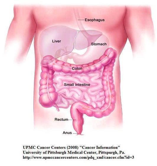
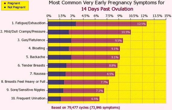
Colon Cancer Stages Early Detection Image
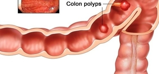
It helps determine how serious the cancer is and how best to treat it. Doctors also use a cancer’s stage when talking about survival statistics. The earliest stage colorectal cancers are called stage 0 (a very early cancer), and then View Diagram Colon Cancer Stages Early Detection Image