Diagram Bones Of Foot Image

This human anatomy diagram with labels depicts and explains the details and or parts of the Diagram Bones Of Foot Image. Human anatomy diagrams and charts show internal organs, body systems, cells, conditions, sickness and symptoms information and/or tips to ensure one lives in good health.

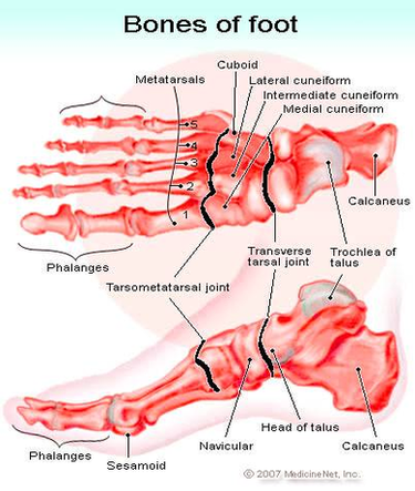
When one looks at the anatomy of the foot, they would realize that the foot has a complex mechanical and structural architecture. The ankle joint is the shock absorber of the foot. Apart from 28 bones, 33 joints, muscles, ligaments, and about 100 foot tendons make the foot. The diagram of bones in the ankle and foot is given below:

The foot is divided into three sections – the forefoot, the midfoot and the hindfoot. The forefoot. This consists of five long metatarsal bones and five shorter bones that form the toes (phalanges). The midfoot. This is approximately pyramid-shaped and is comprised of three cuneiform bones, the navicular bone and the cuboid bone.

The bones of the foot are organized into the tarsal bones, metatarsal bones, and phalanges. The foot begins at the lower end of the tibia and fibula, the two bones of the lower leg. At the base of those, a grouping of bones form the tarsals, which make up the ankle and upper portion of the foot.

Diagram Bones Of Foot Image

Diagram Bones Of Foot Image


Tags: ,