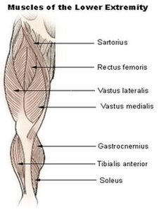
Lower Extremity Diagram Image
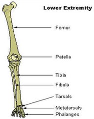
The lower extremity includes the hip, knee, and ankle joints, and the bones of the thigh, leg, and foot. Many people refer to the lower extremity as the leg. In fact, the leg is the part of the body between View Diagram Lower Extremity Diagram Image