Diagram Of Px Human Brain Development Timeline Image

Before we have a look at the brain diagram, it is important to go through a few facts about the brain and its function. This will help you understand the anatomy of the brain better. The average dimension of the adult human brain is 5.5 inches in width and 6.5 inches in length.

The diagram of the brain is useful for both Class 10 and 12. It is one among the few topics having the highest weightage of marks and is frequently asked in the examinations. A well-labelled diagram of a human brain is given below for further reference. The human brain is divided into three main parts:

Let’s review each of the five stages of human brain growth: Stage 1: 0 to 10 months. Neurons and connections growing. Pregnant woman should stay as stress-free as possible, take folic acid, B6 & B12, stimulate this young developing brain with sounds and sensations. Mother should avoid toxins, cigarettes, heavy metals, alcohol, drugs.

Diagram Of Px Human Brain Development Timeline Image Diagram - Chart - diagrams and charts with labels. This diagram depicts Diagram Of Px Human Brain Development Timeline Image

Diagram Of Px Human Brain Development Timeline Image

Diagram Px Flat Stomach Muscles Image

Given below is a labeled diagram of the stomach to help you understand stomach anatomy. The stomach is divided into four parts. These include: Cardia refers to the section of the stomach that is located around the cardiac orifice. The lower esophageal sphincter lies at the junction where the esophagus meets the stomach.

136,936 abdominal muscle stock photos, vectors, and illustrations are available royalty-free.

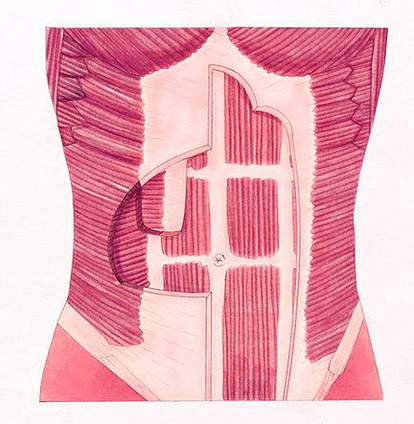
Muscles. The rectus abdominis is the large muscle in the mid-section of the abdomen. It enables the tilt of the pelvis and the curvature of the lower spine. Next to it on both sides of the body is the internal oblique. This wide muscle helps to rotate the spine, increases pressure within the abdomen…

Diagram Px Flat Stomach Muscles Image Diagram - Chart - diagrams and charts with labels. This diagram depicts Diagram Px Flat Stomach Muscles Image

Diagram Px Flat Stomach Muscles Image

Diagram Px Basic Heart Development Timeline Image

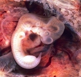
The best place for students to start is the Heart Tutorial. In human embryos the heart begins to beat at about 22-23 days, with blood flow beginning in the 4th week. The heart is therefore one of the earliest differentiating and functioning organs. The heart begins very early in mesoderm within the trilaminar embryonic disc.

Heart development. This begins with the formation of two endocardial tubes which merge to form the tubular heart, also called the primitive heart tube, that loops and septates into the four chambers and paired arterial trunks that form the adult heart. The heart is the first functional organ in vertebrate embryos, and in the human,…

Endocardial tubes. This early development is critical for subsequent embryonic and prenatal development. The heart is the first functional organ to develop and starts to beat and pump blood at around day 21 or 22. The heart begins to develop near the head of the embryo in the cardiogenic area.

Diagram Px Basic Heart Development Timeline Image Diagram - Chart - diagrams and charts with labels. This diagram depicts Diagram Px Basic Heart Development Timeline Image

Diagram Px Basic Heart Development Timeline Image

Diagram Of Px Recognize Prostate Cancer Symptoms Step Image

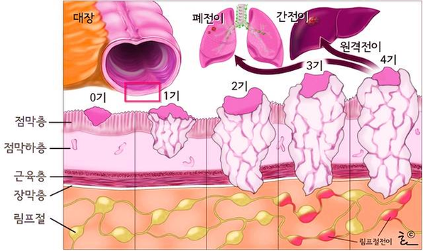
Once the T, N, and M categories have been determined, this information is combined (along with the Grade Group and PSA level if they are available) in a process called stage grouping to get the overall stage of the cancer. The main stages of prostate cancer range from I (1) through IV (4). Some stages are split further (A, B, etc).

This first prostate diagram shows the whole urinary tract system from the kidneys, down through the two ureters into the bladder, through the urethra tube passing inside the prostate gland and out the penis. That is why it sure feels good to go when you gotta go!

Experts say prostate cancer may have no symptoms at first, but you may develop trouble urinating, weak or interrupted urination, frequent urination, trouble emptying your bladder, pain or burning during urination, blood in your urine or semen, painful intercourse, and pain in your back, hips, or pelvis. [2]

Diagram Of Px Recognize Prostate Cancer Symptoms Step Image Diagram - Chart - diagrams and charts with labels. This diagram depicts Diagram Of Px Recognize Prostate Cancer Symptoms Step Image

Diagram Of Px Recognize Prostate Cancer Symptoms Step Image

Px Tubal Pregnancy With Embryo Image

Tubal pregnancy: A pregnancy that is not in the usual place within the uterus but is located in the Fallopian tube. Tubal pregnancies are due to the inability of the fertilized egg to make its way through the Fallopian tube into the uterus.

Tubal pregnancies are most often diagnosed in the sixth through the ninth weeks of pregnancy. 5 Your healthcare provider will be able to diagnose a tubal pregnancy after a medical exam and other testing, which may include: 6 Pelvic examination to test for pain, tenderness, or a mass in the abdomen

Pregnancy tests taken at the beginning of a tubal pregnancy will usually show a positive result, even though the pregnancy won’t be able to grow into a healthy baby. Some people may experience no symptoms at first, but as the fertilized egg continues to grow in the fallopian tube, there can be some serious and more noticeable signs, including: 2

Px Tubal Pregnancy With Embryo Image Diagram - Chart - diagrams and charts with labels. This diagram depicts Px Tubal Pregnancy With Embryo Image

Px Tubal Pregnancy With Embryo Image

Diagram Of Staging Px Image

The method overcomes physical signs of pixel modifications while achieving a high data payload. This technique enables data to be hidden in a cover image, while the image recognition artificial neural network checks the presence of any visible alterations on the stego-image.

Introduction based on its features. Image classification came into existence for by training the computer with the data. The image classification is based on the content of the vision. Motivation by [1], in this paper,

This paper proposed and implemented an image steganography technique (proposed DCT LSB-2) that does not interfere with existing image recognition neural network systems. This technique enables data to be hidden in a cover image, while the image recognition ANN checks the presence of any visible alterations on the stego-image.

Diagram Of Staging Px Image Diagram - Chart - diagrams and charts with labels. This diagram depicts Diagram Of Staging Px Image

Diagram Of Staging Px Image

Px Tell If Dog Is Pregnant Step Image

However, these are also the signs associated with pseudopregnancy, or false pregnancy, where the bitch behaves as if she is pregnant when in fact she isn’t. Some bitches with a false pregnancy even start producing milk, so enlarged mammary glands are not conclusive evidence of pregnancy. Notice if your dog’s nipples are growing.

Stages of Canine Pregnancy Week By Week. Weeks One and Two. An average pregnancy lasts for anything from 56 to 70 days depending on the breed, so this is still early days. Once out of heat, you can continue as usual with your dog – you can groom, feed and walk them exactly as you would if they were not pregnant.

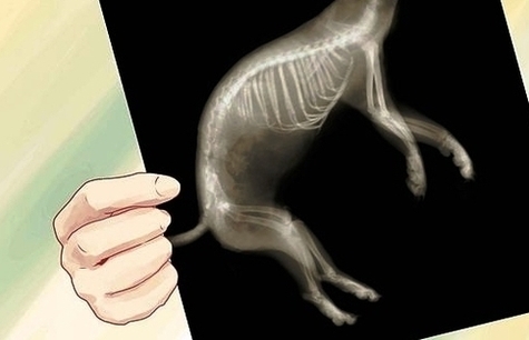
Monitor continuing body changes. In the final third (weeks 6-9) of pregnancy, the dog’s belly becomes rounded and distended. Her mammary glands start to develop and become more obviously swollen, as they get ready to produce milk.

Px Tell If Dog Is Pregnant Step Image Diagram - Chart - diagrams and charts with labels. This diagram depicts Px Tell If Dog Is Pregnant Step Image

Px Tell If Dog Is Pregnant Step Image

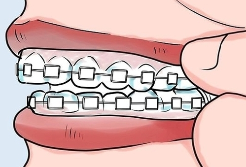
Px Remove Your Own Orthodontic Work Step Image

This means that many of the families that begin an orthodontic treatment move before their treatment has been completed. While getting into orthodontic treatment can be easy, transferring your treatment to a new provider can be a stressful and frustrating process.

OrthoFX clear aligners are used to correct mild-to-moderate orthodontic problems. They are near-invisible and hence, more suitable for those seeking a discreet orthodontic treatment.

For controlled lighting situations where there is generally plenty of light, such as in orthodontic offices, the ISO can remain low to maintain the clarity or sharpness of the image. The ideal ISO for intraoral images would be from 100 to 400 and can be varied based on the lighting conditions in your office.

Px Remove Your Own Orthodontic Work Step Image Diagram - Chart - diagrams and charts with labels. This diagram depicts Px Remove Your Own Orthodontic Work Step Image

Px Remove Your Own Orthodontic Work Step Image