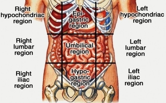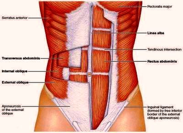
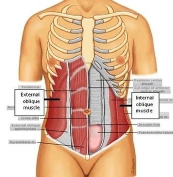
Atlas Tone Abdominal Muscles Image

The Atlas Muscle Toner is clinically demonstrated to deliver firmer, stronger and more toned abdominal muscles while you are: at home, at work, watching TV, exercising, folding laundry, helping your kids with their homework, taking a walk…Doing virtually anything! Atlas View Diagram Atlas Tone Abdominal Muscles Image