Lower Extremity Diagram Image

The lower extremity includes the hip, knee, and ankle joints, and the bones of the thigh, leg, and foot. Many people refer to the lower extremity as the leg. In fact, the leg is the part of the body between the knee and ankle joints. The proper way to describe the lower limb is the lower extremity. This may seem like a minor detail.

The five lower extremity arteries that are routinely examined on ultrasound include the common femoral artery (CFA), the superficial femoral artery (SFA), the popliteal artery, the posterior tibial artery (PTA), and the dorsalis pedis artery (DPA). The common femoral artery (CFA)

This cross-sectional human anatomy atlas of the lower limb is an interactive tool based on MRI axial images of the human leg. Anatomical structures of the lower limb (hip, thigh, knee, leg, ankle and foot) and specific regions (compartment of the lower limb) are visible on dynamic labeled images.

Lower Extremity Diagram Image Diagram - Chart - diagrams and charts with labels. This diagram depicts Lower Extremity Diagram Image
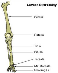
Lower Extremity Diagram Image

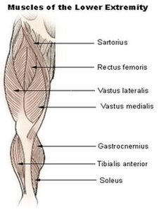
Lower Extremity Muscles Diagram Image

The iliopsoas, an anterior muscle, flexes the thigh. The muscles in the medial compartment adduct the thigh. The illustration below shows some of the muscles of the lower extremity. Muscles that move the leg are located in the thigh region. The quadriceps femoris muscle group straightens the leg at the knee.

When a medical professional refers to your lower extremity, they’re typically referring to everything between your hip to your toes. You lower extremity is a combination of parts: There are over 30 bones in each of your lower extremities including: The muscles in your lower extremity contract and relax to move skeletal bones and thus the body.

The lower leg has two muscle groups to move the ankle, foot, and toes. The bones of the lower leg are the tibia and fibula. The tibia bears most of the weight, and the fibula serves as attachment points for the lower leg muscles. The two tibialis and three peroneus muscles are also found in the lower leg

Lower Extremity Muscles Diagram Image Diagram - Chart - diagrams and charts with labels. This diagram depicts Lower Extremity Muscles Diagram Image

Lower Extremity Muscles Diagram Image

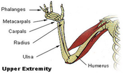
Upper Extremity Diagram1 Image

The forearm is the portion between the elbow and wrist. The thigh is the portion of the lower extremity between the hip and knee, and the calf is the portion between the knee and ankle. The normal arterial anatomy of the upper extremity is depicted graphically in Figure 13-1.

The next chapter is about the osteology of the upper limb, with all its anatomical structures, muscle insertions and ligaments of the bones: scapula, clavicle, humerus, ulna, radius, carpal bones, metacarpals and phalanges of the fingers.

The final chapter presents anatomical charts of anatomical sections of the upper limb: the axilla and the deltoid region in axial and coronal and axial sections of the arm, the elbow, forearm, wrist, carpal and metacarpal regions.

Upper Extremity Diagram1 Image Diagram - Chart - diagrams and charts with labels. This diagram depicts Upper Extremity Diagram1 Image

Upper Extremity Diagram1 Image

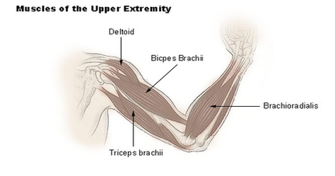
Upper Extremity Muscles Diagram Image

Upper limb muscles and movements. The upper limb (upper extremity) is truly a complex part of human anatomy. It is best studied broken down into its components: regions, joints, muscles, nerves, and blood vessels.

Upper limb muscles and movements. 1 Scapular region. The scapular region lies on the posterior surface of the thoracic wall. It may seem strange that it is included in the anatomy of the … 2 Shoulder. 3 Arm (brachium) 4 Forearm flexors. 5 Forearm extensors. More items

The upper arm is located between the shoulder joint and elbow joint. It contains four muscles – three in the anterior compartment (biceps brachii, brachialis, coracobrachialis), and one in the posterior compartment (triceps brachii). In this article, we shall look at the anatomy of the muscles…

Upper Extremity Muscles Diagram Image Diagram - Chart - diagrams and charts with labels. This diagram depicts Upper Extremity Muscles Diagram Image

Upper Extremity Muscles Diagram Image

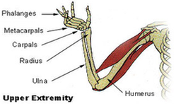
Upper Extremity Diagram Image

Anatomy of the upper extremity in standard radiology This radioanatomy module on the upper limb presents 16 radiographic images with 112 structures labeled.

The forearm is the portion between the elbow and wrist. The thigh is the portion of the lower extremity between the hip and knee, and the calf is the portion between the knee and ankle. The normal arterial anatomy of the upper extremity is depicted graphically in Figure 13-1.

Plain X-Rays of the upper limb The first X-Ray image is focused on the pectoral girdle seen from the front, allowing us to study the clavicles, sternum and sternoclavicular joints. The following image shows the clavicle in an AP view, with its sternal and acromial extremities, conoid tubercle and the body of the clavicle.

Upper Extremity Diagram Image Diagram - Chart - diagrams and charts with labels. This diagram depicts Upper Extremity Diagram Image

Upper Extremity Diagram Image