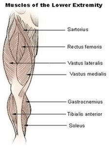
Lower Extremity Muscles Diagram Image

This human anatomy diagram with labels depicts and explains the details and or parts of the Lower Extremity Muscles Diagram Image. Human anatomy diagrams and charts show internal organs, body systems, cells, conditions, sickness and symptoms information and/or tips to ensure one lives in good health.

The iliopsoas, an anterior muscle, flexes the thigh. The muscles in the medial compartment adduct the thigh. The illustration below shows some of the muscles of the lower extremity. Muscles that move the leg are located in the thigh region. The quadriceps femoris muscle group straightens the leg at the knee.

When a medical professional refers to your lower extremity, they’re typically referring to everything between your hip to your toes. You lower extremity is a combination of parts: There are over 30 bones in each of your lower extremities including: The muscles in your lower extremity contract and relax to move skeletal bones and thus the body.

The lower leg has two muscle groups to move the ankle, foot, and toes. The bones of the lower leg are the tibia and fibula. The tibia bears most of the weight, and the fibula serves as attachment points for the lower leg muscles. The two tibialis and three peroneus muscles are also found in the lower leg

Lower Extremity Muscles Diagram Image

Lower Extremity Muscles Diagram Image


Tags: , ,