Diagram Px Flat Stomach Muscles Image

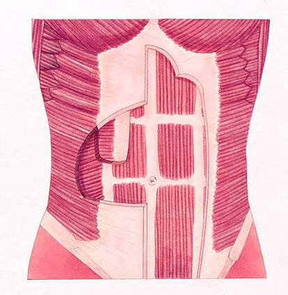
Given below is a labeled diagram of the stomach to help you understand stomach anatomy. The stomach is divided into four parts. These include: Cardia refers to the section of the stomach that is located around the cardiac orifice. The lower esophageal sphincter lies at the junction where the esophagus meets the stomach.

136,936 abdominal muscle stock photos, vectors, and illustrations are available royalty-free.

Muscles. The rectus abdominis is the large muscle in the mid-section of the abdomen. It enables the tilt of the pelvis and the curvature of the lower spine. Next to it on both sides of the body is the internal oblique. This wide muscle helps to rotate the spine, increases pressure within the abdomen…

Diagram Px Flat Stomach Muscles Image Diagram - Chart - diagrams and charts with labels. This diagram depicts Diagram Px Flat Stomach Muscles Image

Diagram Px Flat Stomach Muscles Image

Food Pyramid Flat Sm Image

475 food pyramid stock photos and images available, or search for food pyramid vector or healthy food pyramid to find more great stock photos and pictures.

The food pyramid infographic with food icons and categories, health tips at bottom Table filled with different types of foods A large group of food. The assortment includes fruits, vegetables, meat, fish and dairy products. The food is mostly sorted by type.

The USDA “ChooseMyPlate” basic food group for healthy diet, with a plate of food choices including grains, protein, vegetables, fruits and dairy. This plate pie chart concept replaces the former food pyramid recommendaton for guidance on the amount of milk, bread, pasta, chicken and other foods advised for balanced meals. Food pyramid.

Food Pyramid Flat Sm Image Diagram - Chart - diagrams and charts with labels. This diagram depicts Food Pyramid Flat Sm Image

Food Pyramid Flat Sm Image

Dorsal Forearm Diag Flat Image

Anatomy of the distal forearm 1 Soft tissue anatomy – dorsal. 2 Soft tissue anatomy – palmar. 3 Bony anatomy. 4 Principle of columns. The distal forearm may be thought of in terms of three columns. The ulna forms one column. The… More …

Currently, computed tomography (CT) and magnetic resonance imaging (MRI) play an important role in evaluation of injuries to the distal forearm, wrist, and hand (see the text that follows).

Volar/dorsal splints are splints that extend from the middle of the forearm to the distal palmar crease (top crease in the palm). Volar splints are applied to the palm side of the forearm, wrist and hand, while dorsal splints are applied to the top side of the forearm, wrist, and hand.

Dorsal Forearm Diag Flat Image Diagram - Chart - diagrams and charts with labels. This diagram depicts Dorsal Forearm Diag Flat Image

Dorsal Forearm Diag Flat Image