Diagram Anatomy Ear Image

This human anatomy diagram with labels depicts and explains the details and or parts of the Diagram Anatomy Ear Image. Human anatomy diagrams and charts show internal organs, body systems, cells, conditions, sickness and symptoms information and/or tips to ensure one lives in good health.

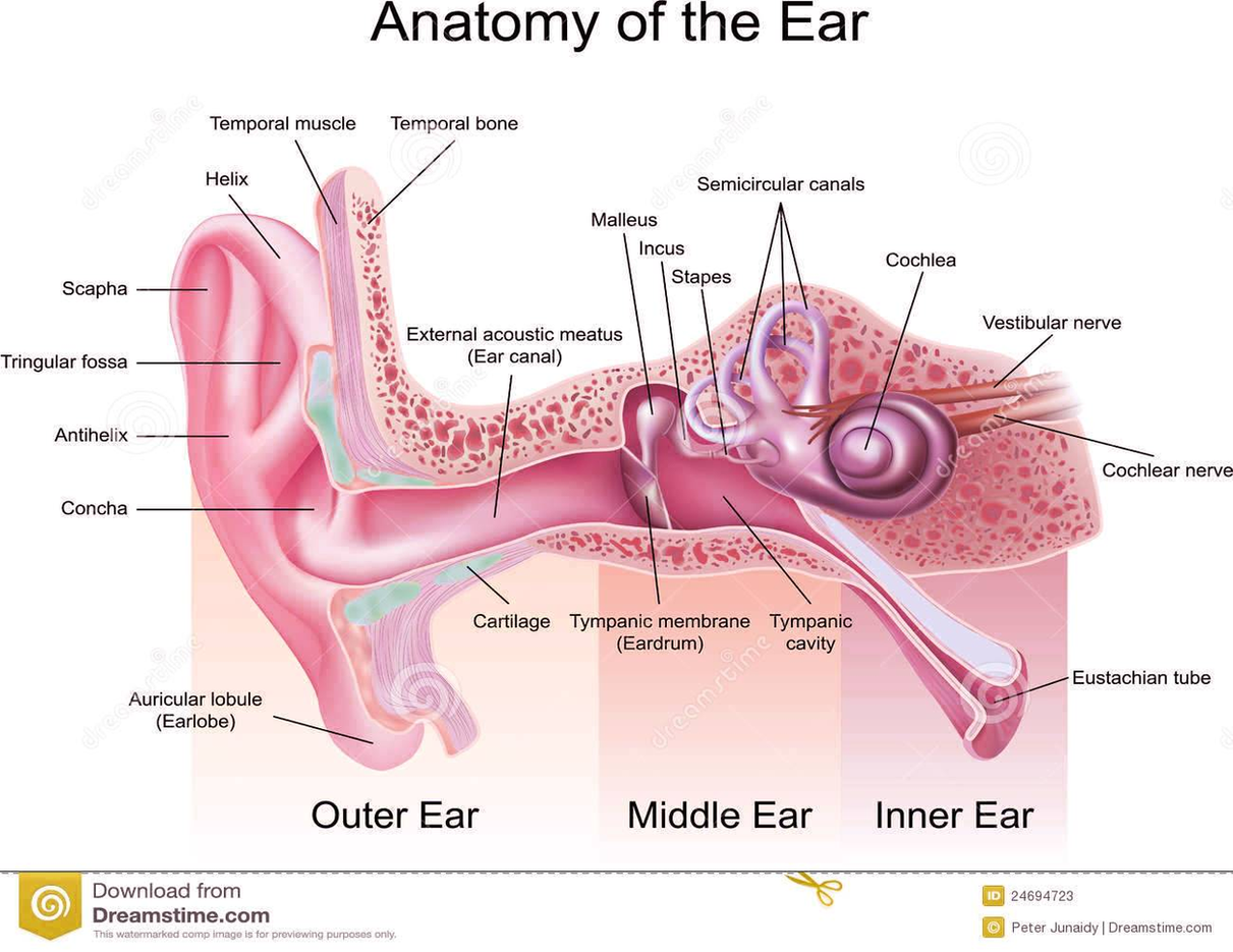
The Structure of Human Ear. Helix: It is the prominent outer rim of the external ear. Antihelix: It is the cartilage curve that is situated parallel to the helix. Crus of the Helix: It is the landmark of the outer ear, situated right above the pointy protrusion known as the tragus.

Ear Anatomy Images. The ear drum is often transparent and looks like a stretched piece of clear plastic. The drum is approximately the size of a dime, with the newborn ear drum the same size as the adult. The malleus is the middle ear bone which is attached to the drum and easily identified.

The drum looks healthy and has a nice gray color to it. The image is of a normal nasopharynx and the opening to the Eustachian tube. The Eustachian tube goes from the back of the nose (nasopharynx) to the middle ear. Normally the tube remains closed and opens when you swallow, yell or pop your ear with a Valsalva Maneuver.

Diagram Anatomy Ear Image

Diagram Anatomy Ear Image


Tags: