Diagram Of Nasa Middle Ear Image

This human anatomy diagram with labels depicts and explains the details and or parts of the Diagram Of Nasa Middle Ear Image. Human anatomy diagrams and charts show internal organs, body systems, cells, conditions, sickness and symptoms information and/or tips to ensure one lives in good health.

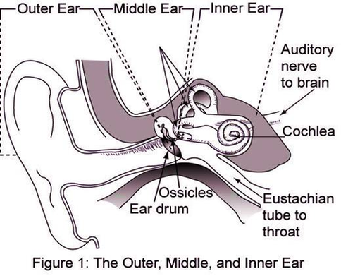
Also known as the tympanic cavity, the middle ear is an air-filled, membrane-lined space located between the ear canal and the Eustachian tube, cochlea, and auditory nerve. The eardrum separates this space from the ear canal.

The Structure of Human Ear. Helix: It is the prominent outer rim of the external ear. Antihelix: It is the cartilage curve that is situated parallel to the helix. Crus of the Helix: It is the landmark of the outer ear, situated right above the pointy protrusion known as the tragus.

The Structure of Human Ear Helix: It is the prominent outer rim of the external ear. Antihelix: It is the cartilage curve that is situated parallel to the helix. Crus of the Helix: It is the landmark of the outer ear, situated right above the pointy protrusion known as the tragus.

Diagram Of Nasa Middle Ear Image

Diagram Of Nasa Middle Ear Image


Tags: , ,