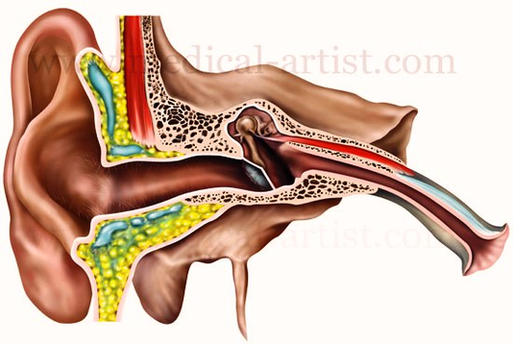
Ear Anatomy Illustration Image

This human anatomy diagram with labels depicts and explains the details and or parts of the Ear Anatomy Illustration Image. Human anatomy diagrams and charts show internal organs, body systems, cells, conditions, sickness and symptoms information and/or tips to ensure one lives in good health.

12,612 human ear anatomy stock photos, vectors, and illustrations are available royalty-free.

In most mammals, the visible ear is a flap of tissue that is also called the pinna or the auricle. Vertebrates have a pair of ears, placed somewhat symmetrically on opposite sides of the head. This arrangement aids in the ability to localise sound sources.

There are three components to the ear: the outer ear, the middle ear and the inner ear. All three are involved in hearing but only the inner ear is responsible for balance. In most mammals, the visible ear is a flap of tissue that is also called the pinna or the auricle.

Ear Anatomy Illustration Image

Ear Anatomy Illustration Image


Tags: ,