Eye Muscles Diagram

This human anatomy diagram with labels depicts and explains the details and or parts of the Eye Muscles Diagram. Human anatomy diagrams and charts show internal organs, body systems, cells, conditions, sickness and symptoms information and/or tips to ensure one lives in good health.

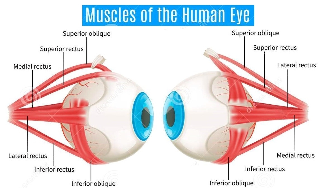
Eye muscles. There are two groups of eye muscles: Six extraocular muscles move the eye: superior rectus, inferior rectus, medial rectus, lateral rectus, superior oblique and inferior oblique muscles; and one other, levator palpebrae superioris, opens the eyelid.

Intrinsic ocular muscles which are within the eyeball itself and control how the eyes accommodate Six extraocular muscles move the eye: superior rectus, inferior rectus, medial rectus, lateral rectus, superior oblique and inferior oblique muscles; and one other, levator palpebrae superioris, opens the eyelid.

There are four recti muscles; superior rectus, inferior rectus, medial rectus and lateral rectus. These muscles characteristically originate from the common tendinous ring. This is a ring of fibrous tissue, which surrounds the optic canal at the back of the orbit.

Eye Muscles Diagram

Eye muscles diagram


Tags: , , ,