Human Pelvis Image Human male anatomy scheme. Main pelvic bones – sacrum, ilium, coccyx, pubis, ischium. Vector illustration isolated on a white background. pelvis anatomy stock illustrations Human male anatomy scheme. Main pelvic bones – sacrum, ilium, coccyx, pubis, ischium. Vector illustration isolated on a white background.
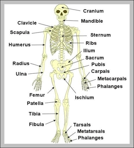
Evolutionary scientists believe this stems from man’s hunter roots, as a leaner pelvis made running easier. The bones of the pelvis are the hip bones, sacrum, and coccyx. Each hip bone contains three bones — the ilium, ischium, and pubis — that fuse together as we grow older.
The pelvic region is the area between the trunk — or main body — and the lower extremities, or legs. The male pelvis is different from a female’s. The pelvic bones are smaller and narrower. Evolutionary scientists believe this stems from man’s hunter roots, as a leaner pelvis made running easier.
Skeletal System Pelvis Image Diagram - Chart - diagrams and charts with labels. This diagram depicts Skeletal System Pelvis Image