Calf Muscle Diagram Image

This human anatomy diagram with labels depicts and explains the details and or parts of the Calf Muscle Diagram Image. Human anatomy diagrams and charts show internal organs, body systems, cells, conditions, sickness and symptoms information and/or tips to ensure one lives in good health.

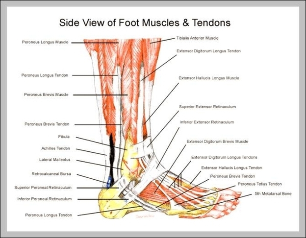
The calf is defined largely as the three headed calf muscle (triceps surae muscle). In anatomy, we actually talk about the lower leg muscles and divide them into the following categories:

Picture of the Calf Muscle. The gastrocnemius is the larger calf muscle, forming the bulge visible beneath the skin. The gastrocnemius has two parts or “heads,” which together create its diamond shape. The soleus is a smaller, flat muscle that lies underneath the gastrocnemius muscle.

Calf muscle ultrasound: A device placed on the skin bounces high-frequency sound waves off the calf muscle, tendon, and other structures in the leg. The signals are converted into images on a video screen, allowing health-care providers to see structures inside the body. 1 2

Calf Muscle Diagram Image

Calf Muscle Diagram Image


Tags: ,