Distal Aspect of the Humerus Epicondyle Trochlea Capitulum
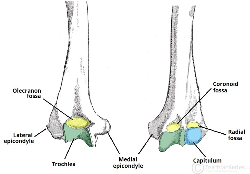
The distal humerus features several important bony landmarks, including the medial and lateral epicondyles, the trochlea, and the capitulum, all of which play key roles in elbow function. The epicondyles serve as attachment points for the forearms flexor and extensor View Diagram Distal Aspect of the Humerus Epicondyle Trochlea Capitulum