Final Long Bone Diagram Image

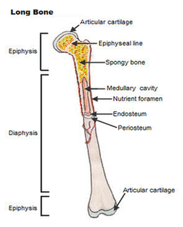
Long bone anatomy A long bone is a bone that has greater length than width. A long bone has a shaft and 2 ends. Long bones have a thick outside layer of compact bone and an inner medullary cavity containing bone marrow.

Long bone fracture 1 The location of the fracture (the tibial shaft is divided into thirds: distal, middle, and proximal) 2 The pattern of the fracture (for example, the bone can break in different directions, such as crosswise, lengthwise, or in the middle) 3 Whether the skin and muscle over the bone is torn by the injury (open fracture)

Inside the diaphysis is the medullary cavity, which is filled with yellow bone marrow in an adult. The outer walls of the diaphysis (cortex, cortical bone) are composed of dense and hard compact bone, a form of osseous tissue. Figure 6.3.1 – Anatomy of a Long Bone: A typical long bone showing gross anatomical features.

Final Long Bone Diagram Image Diagram - Chart - diagrams and charts with labels. This diagram depicts Final Long Bone Diagram Image

Final Long Bone Diagram Image