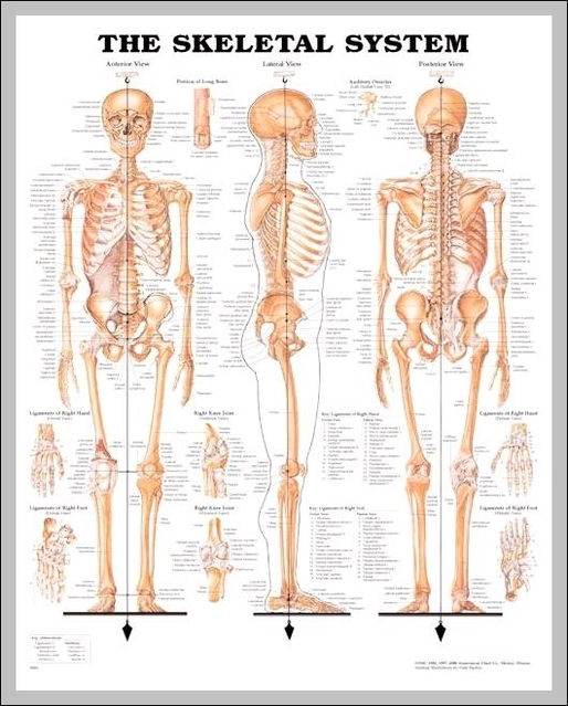
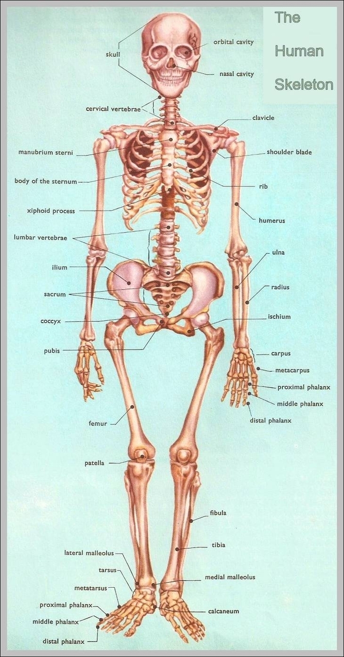
Picture Of Human Vertebrae
The human spine is composed of 33 vertebrae that interlock with each other to form the spinal column. The human spinal cord consists of nerves that connect the brain to nerves in the body. The vertebrae of the spine align View Diagram Picture Of Human Vertebrae