Picture Of a Human Kidney

This human anatomy diagram with labels depicts and explains the details and or parts of the Picture Of a Human Kidney. Human anatomy diagrams and charts show internal organs, body systems, cells, conditions, sickness and symptoms information and/or tips to ensure one lives in good health.

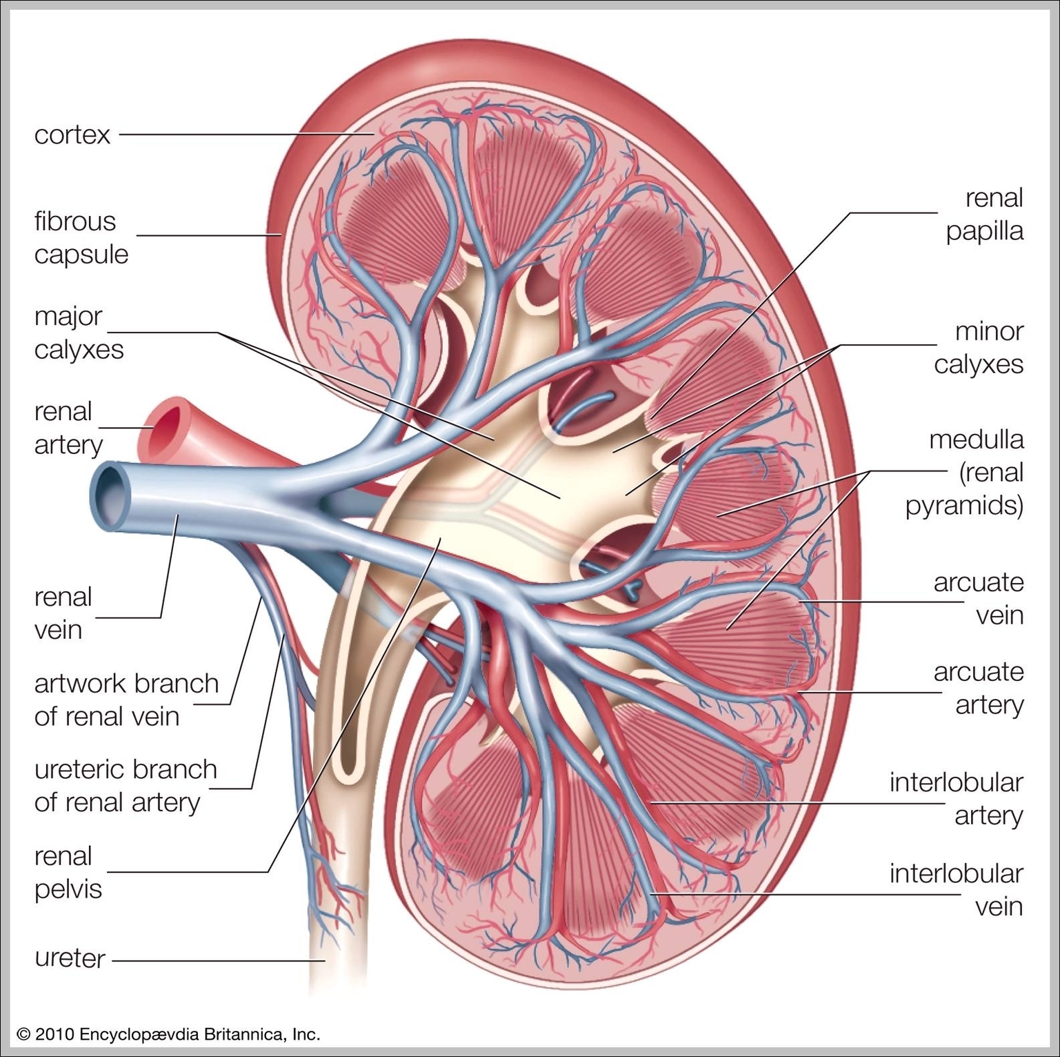
The kidneys are two organs that lie on either side of the spine just under the rib cage. Kidneys serve many important functions including filtering blood, controlling electrolyte balance, and producing hormones.

Computed tomography (CT) scan: A CT scanner takes a series of X-rays, and a computer creates detailed images of the kidneys. Magnetic resonance imaging (MRI) scan: A scanner uses radio waves in a magnetic field to make high-resolution images of the kidneys.

Picture of Kidneys. Nephrons are the small filtering units in the kidneys. Nephrons filter wastes and excess fluid out of the blood. Concentrated waste (urine) travels from the kidneys down tubes called the ureters and into the bladder. The kidneys produce approximately 1 to 2 quarts of urine per day.

Picture Of a Human Kidney

picture of a human kidney


Tags: , , , , , , ,