
Stomach Anatomy Diagram
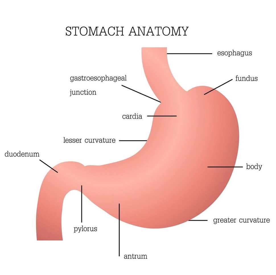
Stomach Anatomy Diagram: A stomach diagram shows its regionscardia, fundus, body, and pylorusas well as its inner lining and muscular layers that aid in digestion.
Stomach Anatomy Diagram: A stomach diagram shows its regionscardia, fundus, body, and pylorusas well as its inner lining and muscular layers that aid in digestion.
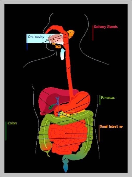
The stomach is an important organ in the digestive system. After food has been chewed in the mouth and swallowed, it enters the stomach via the oesophagus. The stomach produces strong acid. This kills many harmful microorganisms that might have View Diagram What Does The Stomach Do
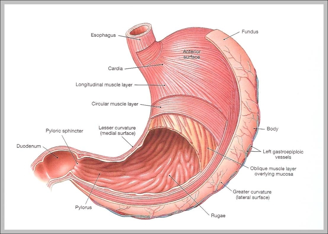
Stomach. When the stomach is empty, the inside has small folds called rugae. Rugae allow the stomach to expand to accommodate large meals. They also grip the food inside the stomach to help physically break it down. The average stomach View Diagram Picture Of a Stomach

Abdominal muscle, any of the muscles of the anterolateral walls of the abdominal cavity, composed of three flat muscular sheets, from without inward: external oblique, internal oblique, and transverse abdominis, supplemented in front on each side of the midline by View Diagram Stomach Muscle Anatomy
The enzymes in the mouth and stomach include amylase, lipase and pepsin — and each is responsible for helping to start the digestion process for carbohydrates, fat and protein. It Starts in the Mouth The function of stomach enzymes is View Diagram Stomach Enzymes
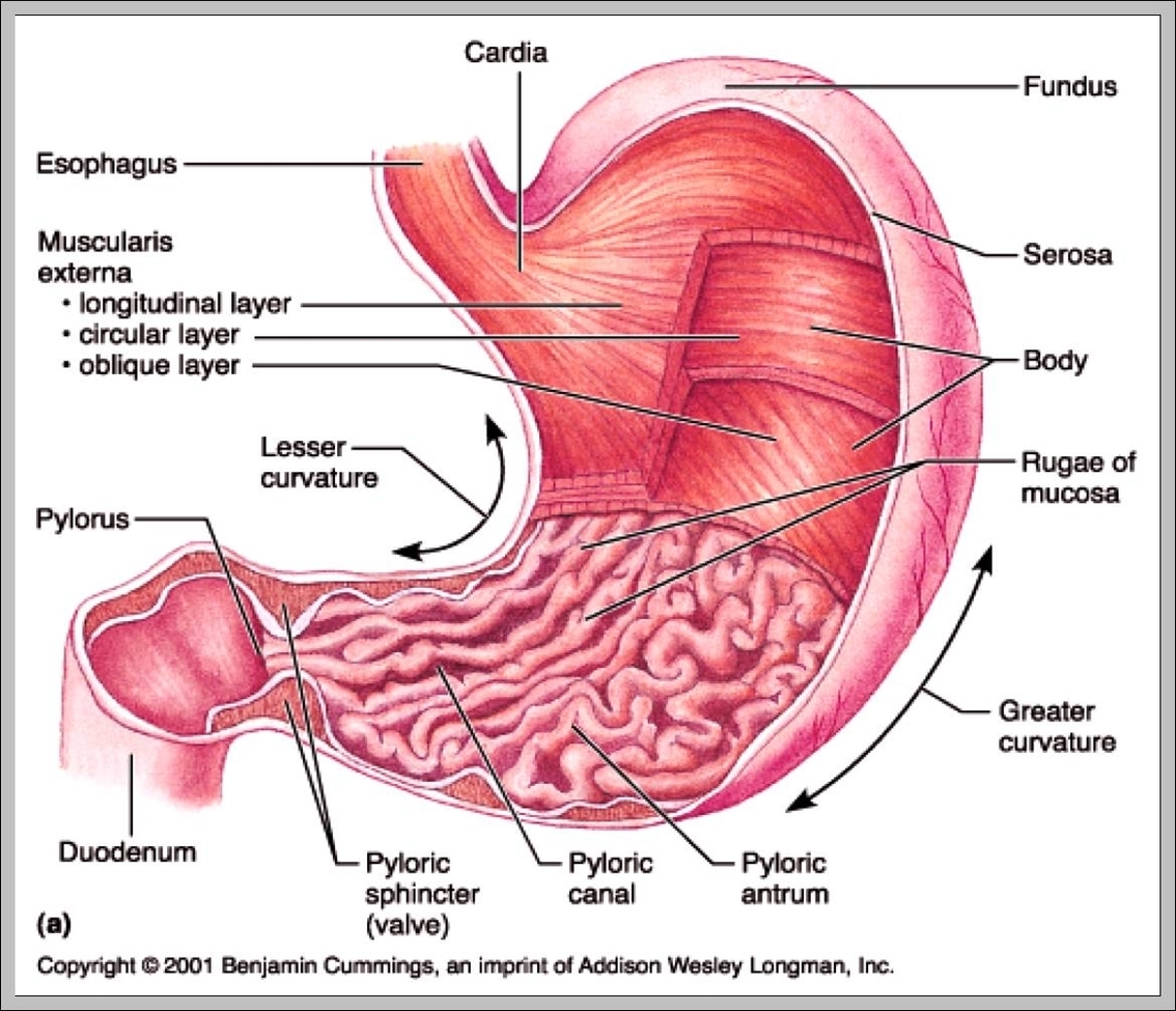
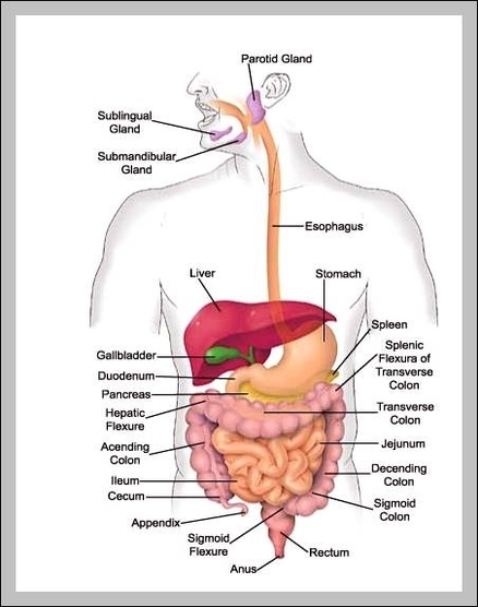
Stomach Location (Anatomical Position), Parts and Pictures. The stomach is a hollow organ that lies between the esophagus (food pipe) and duodenum (small intestine). It is an expanded part of the gastrointestinal tract (gut) that plays an important role in View Diagram Stomach Location
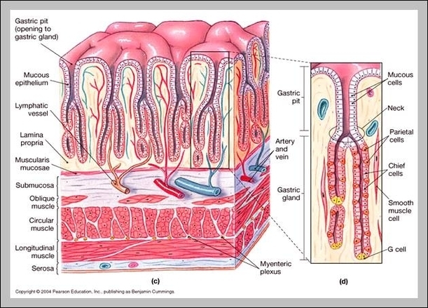
A number of diseases can involve the stomach lining. Like other cells, the epithelial cells in the stomach can become cancerous, causing the development of stomach cancers. The stomach lining can also become inflamed in a condition known as gastritis, View Diagram Stomach Lining
Stomach Cells: The stomach contains several specialized cellsparietal, chief, and mucous cellsthat produce gastric acid, enzymes, and protective mucus to aid in digestion and prevent tissue damage.