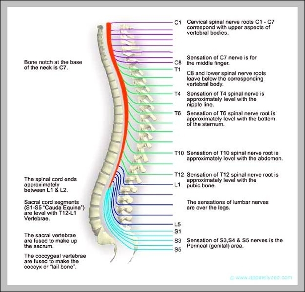
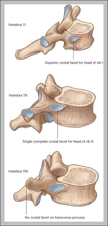
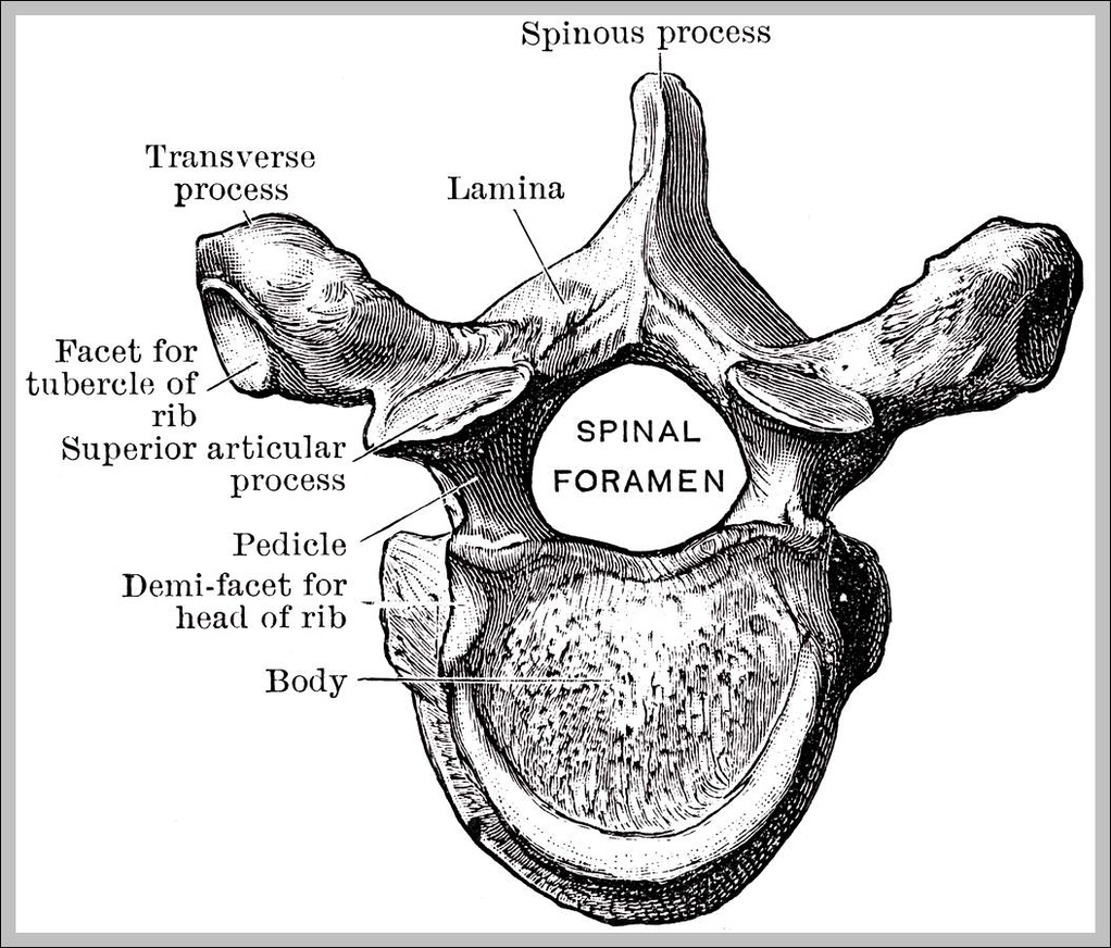
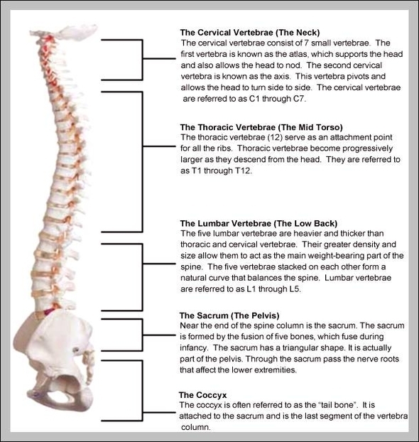
T 11 Vertebrae

T11 is an atypical thoracic vertebra. In contrast to typical thoracic vertebrae, it contains a single costal facet that articulates with the atypical eleventh rib.

The eleventh thoracic vertebra (T11) is located near the bottom of the thoracic spine. Generally, the spinal bones are larger and heavier the further down they are located. This helps each vertebra to support the weight of the vertebrae above it, as well as the weight of the skull and brain.

Some common symptomatic signs of T11 or T12 subluxations (misalignments) are things like blood sugar problems, kidney conditions, gas pains, chronic tiredness, constipation, diarrhea, lower rib pain or sciatica. Now, we all know there are a plethora of different causes for each of these symptoms!

T 11 Vertebrae Diagram - Chart - diagrams and charts with labels. This diagram depicts T 11 Vertebrae

t 11 vertebrae

T 7 Vertebrae

Thoracic Vertebrae and Nerve Interactions at T7 *. Subluxation at this area can result in gastritis and ulcers. T7 is in the middle of the twelve vertebrae of the torso section of the spinal column. It has a strong center section and is larger than T6 and smaller than T8. The foramen at this vertebrae are smaller than ones at T6.

As the transitional vertebra between the cervical and thoracic regions, the C7 has some features of both the cervical and thoracic vertebrae. Like the C3 through C6 vertebrae, C7 has a thicker area of bone called the body, located anterior to the vertebral foramen.

Continued From Above… The anterior portion of the T7 vertebra is made of a wide, heart-shaped cylinder of bone known as the centrum or vertebral body. The centrum provides most of the strength to the vertebra and connects it to the surrounding vertebrae via the intervertebral disks.

T 7 Vertebrae Diagram - Chart - diagrams and charts with labels. This diagram depicts T 7 Vertebrae

t 7 vertebrae

Vertebrae In Neck

The vertebrae at the top of the neck tend to be smaller and more mobile while the lower cervical vertebrae are larger to handle greater loads from the neck and head above. C3, C4, C5, and C6 cervical vertebrae.

The neck is connected to the upper back through a series of seven vertebral segments. The cervical spine has 7 stacked bones called vertebrae, labeled C1 through C7. The top of the cervical spine connects to the skull, and the bottom connects to the upper back at about shoulder level.

A fracture (break or crack) in one of the seven cervical vertebrae in your neck that support your head and connect it to the shoulders and body is called a cervical fracture or sometimes, a broken neck. A broken neck is a big deal because it is the central nervous system’s main, some may say only, connection between the brain and the body.

Vertebrae In Neck Diagram - Chart - diagrams and charts with labels. This diagram depicts Vertebrae In Neck

vertebrae in neck

Picture Of Vertebrae

The lumbar vertebrae consist of five individual cylindrical bones that form the spine in the lower back. These vertebrae carry all of the upper body’s weight while providing flexibility and movement to the trunk region.

A unique feature of the thoracic vertebrae is that each one forms joints with a pair of ribs to form the sturdy rib cage that protects the organs of the chest. Lumbar: The 5 vertebrae in the lower back form the lumbar region of the spine.

The vertebrae of the spine align so that their vertebral canals form a hollow, bony tube to protect the spinal cord from external damage and infection. Between the vertebrae are small spaces known as intervertebral canals that allow spinal nerves to exit the spinal cord and connect to the various regions of the body.

Picture Of Vertebrae Diagram - Chart - diagrams and charts with labels. This diagram depicts Picture Of Vertebrae

picture of vertebrae

Vertebrae In The Neck

The neck is connected to the upper back through a series of seven vertebral segments. The cervical spine has 7 stacked bones called vertebrae, labeled C1 through C7. The top of the cervical spine connects to the skull, and the bottom connects to the upper back at about shoulder level.

· Longus Colli- Begins between the third and sixth cervical vertebrae, responsible for flexion of the head and neck. · Longus Capitis- Begins between the third and sixth cervical vertebrae, responsible for flexion of the neck · Rectus Capitis Anterior- Begins at the first cervical vertebrae, responsible for flexion of the neck

Truncal vertebrae (divided into thoracic and lumbar vertebrae in mammals) lie caudal (toward the tail) of cervical vertebrae. In sauropsid species, the cervical vertebrae bear cervical ribs. In lizards and saurischian dinosaurs, the cervical ribs are large; in birds, they are small and completely fused to the vertebrae.

Vertebrae In The Neck Diagram - Chart - diagrams and charts with labels. This diagram depicts Vertebrae In The Neck

vertebrae in the neck

Pictures Of Vertebrae

A unique feature of the thoracic vertebrae is that each one forms joints with a pair of ribs to form the sturdy rib cage that protects the organs of the chest. Lumbar: The 5 vertebrae in the lower back form the lumbar region of the spine.

The lumbar vertebrae consist of five individual cylindrical bones that form the spine in the lower back. These vertebrae carry all of the upper body’s weight while providing flexibility and movement to the trunk region.

The vertebrae of the spine align so that their vertebral canals form a hollow, bony tube to protect the spinal cord from external damage and infection. Between the vertebrae are small spaces known as intervertebral canals that allow spinal nerves to exit the spinal cord and connect to the various regions of the body.

Pictures Of Vertebrae Diagram - Chart - diagrams and charts with labels. This diagram depicts Pictures Of Vertebrae

pictures of vertebrae

Picture Of Human Vertebrae

The human spine is composed of 33 vertebrae that interlock with each other to form the spinal column. The human spinal cord consists of nerves that connect the brain to nerves in the body.

The vertebrae of the spine align so that their vertebral canals form a hollow, bony tube to protect the spinal cord from external damage and infection. Between the vertebrae are small spaces known as intervertebral canals that allow spinal nerves to exit the spinal cord and connect to the various regions of the body.

A cylinder of bone known as the vertebral body makes up the majority of the lumbar vertebrae’s mass and bears most of the body’s weight. Posteriorly the body is connected to a thin ring of bone known as the arch.

Picture Of Human Vertebrae Diagram - Chart - diagrams and charts with labels. This diagram depicts Picture Of Human Vertebrae

picture of human vertebrae

T5 Vertebrae

The 12 thoracic vertebrae are labeled T1 through T12, with T1 being closest to the skull and T12 being closest to the tailbone. The T5 is the fifth thoracic vertebra closest to the skull. Most thoracic vertebrae have a variety of characteristics in common. The middle portions of these vertebrae are heart-shaped.

The transverse processes are long, with the upper vertebral notches deeper than any of those found on the other thoracic vertebrae. The thoracic spinal nerve 1 passes through underneath T1. Second thoracic vertebrae (T2): The T2 vertebra is larger than T1 vertebra. The thoracic spinal nerve 2 passes through underneath T2.

Your T5 vertebra is located in your spine, roughly halfway down your back. It’s the fifth of our twelve thoracic vertebrae, and is attached to ribs on both sides of your body.

T5 Vertebrae Diagram - Chart - diagrams and charts with labels. This diagram depicts T5 Vertebrae

t5 vertebrae

Thoracic Vertebrae Numbers

The thoracic spine is comprised of 12 vertebrae labeled T1 through T12. The top thoracic vertebra, T1, connects with C7 in the cervical spine above while the bottom thoracic vertebra, T12, connects with L1 in the lumbar spine below. In addition to being connected to adjacent vertebrae, the thoracic vertebrae are also connected to ribs.

Distinguishing features. The body of a thoracic vertebra is somewhat “heart-shaped,” and is larger than the cervical but smaller than the lumbar vertebrae in size. The body also has small, smooth, and somewhat concave costal facets for the attachment of the ribs. Ribs are generally inserted between two vertebrae,…

[edit on Wikidata] In vertebrates, thoracic vertebrae compose the middle segment of the vertebral column, between the cervical vertebrae and the lumbar vertebrae.

Thoracic Vertebrae Numbers Diagram - Chart - diagrams and charts with labels. This diagram depicts Thoracic Vertebrae Numbers

thoracic vertebrae numbers

T 1 Vertebrae

The T1 vertebra is the first (uppermost) of the twelve (12) thoracic vertebrae that make up the central and largest section of the spinal column between the lumbar vertebrae below and the cervical vertebrae above. While larger than the C7 vertebra above it, the T1 is the smallest of the thoracic vertebrae.

The T1 vertebra is the first (uppermost) of the twelve (12) thoracic vertebrae that make up the central and largest section of the spinal column between the lumbar vertebrae below and the cervical vertebrae above. While larger than the C7 vertebra above it, the T1 is the smallest of the thoracic vertebrae.

Individuals who suffered a minor accident or subluxation are more likely to experience less severe injuries like the ones listed below. Here is a list of the T1 through T12 vertebrae and their potential corresponding injuries. Allergies and hives. Ulcers and gastritis.

T 1 Vertebrae Diagram - Chart - diagrams and charts with labels. This diagram depicts T 1 Vertebrae

t 1 vertebrae

Spinal Cord Vertebrae

The spinal cord runs inside the vertebral column, and vertebral column protects the spinal cord. This is the key difference between spinal cord and vertebral column. Spinal cord and vertebral column are two vital structures of humans. They run together from head to abdomen but work independently.

The spinal cord runs inside the vertebral column, and vertebral column protects the spinal cord. This is the key difference between spinal cord and vertebral column. Spinal cord and vertebral column are two vital structures of humans. They run together from head to abdomen but work independently. 1. Overview and Key Difference 2.

The spinal cord is long, thin tubular bundles of nerves composed of 31 pairs. It runs inside the vertebral column, a bony structure with 26 vertebrae. The latter protects the spinal cord and provides attachment sites for ribs and neck muscles. The spinal cord passes information from the brain to peripheral nervous system.

Spinal Cord Vertebrae Diagram - Chart - diagrams and charts with labels. This diagram depicts Spinal Cord Vertebrae

spinal cord vertebrae

T4 Vertebrae

The vertebrae that make up the spinal column have a specific shape that is made to protect the spinal cord from injury. The T4 vertebra is located about midway up the spine. The portion of the spinal cord that the thoracic vertebrae protect is referred to as the thoracic cord. This part of the cord sends and receives signals from the area…

T4 Syndrome: Signs, Symptoms, Causes, Treatment, Recovery. What Is T4 Syndrome Or Upper Thoracic Syndrome? T4 syndrome, also known as Upper Thoracic Syndrome refers to the pain in the upper back and is a much under-recognized or diagnosed condition. The spinal cord is divided into 5 segments: Cervical, thoracic, lumbar, sacral and coccyx.

The T4 vertebra is located about midway up the spine. The portion of the spinal cord that the thoracic vertebrae protect is referred to as the thoracic cord.

T4 Vertebrae Diagram - Chart - diagrams and charts with labels. This diagram depicts T4 Vertebrae

t4 vertebrae

Spinal Vertebrae Chart

These regions are called the cervical spine, thoracic spine, lumbar spine, sacrum and coccyx. There are seven cervical vertebrae, twelve thoracic vertebrae and five lumbar vertebrae. The number of vertebrae in a region can vary but overall the number remains the same.

The vertebral arch is posterior, meaning it faces the back of a person. Together, these enclose the vertebral foramen, which contains the spinal cord. Because the spinal cord ends in the lumbar spine, and the sacrum and coccyx are fused, they do not contain a central foramen.

The second cervical vertebra, C2, is also known as the “axis” because it allows the skull and atlas to rotate to the left and right. Thoracic: The 12 vertebrae in the chest region form the spine’s thoracic region. Thoracic vertebrae are larger and stronger than cervical vertebrae but are much less flexible.

Spinal Vertebrae Chart Diagram - Chart - diagrams and charts with labels. This diagram depicts Spinal Vertebrae Chart

spinal vertebrae chart